. 2013 Jun;87(11):6182-91.
doi: 10.1128/JVI.03024-12. Epub 2013 Mar 27.

Tyrosine 132 phosphorylation of influenza A virus M1 protein is crucial for virus replication by controlling the nuclear import of M1

Affiliations

Tyrosine 132 phosphorylation of influenza A virus M1 protein is crucial for virus replication by controlling the nuclear import of M1

Shanshan Wang et al. J Virol. 2013 Jun.

Abstract

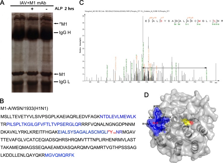
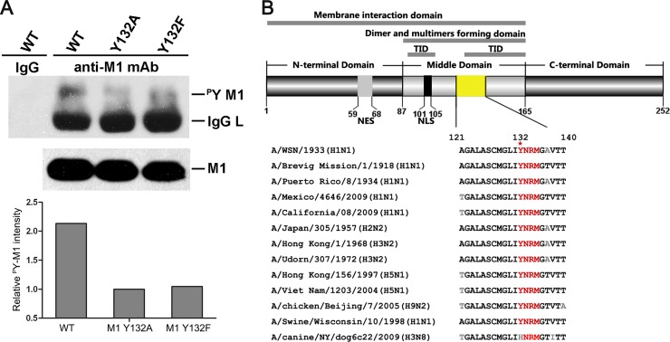
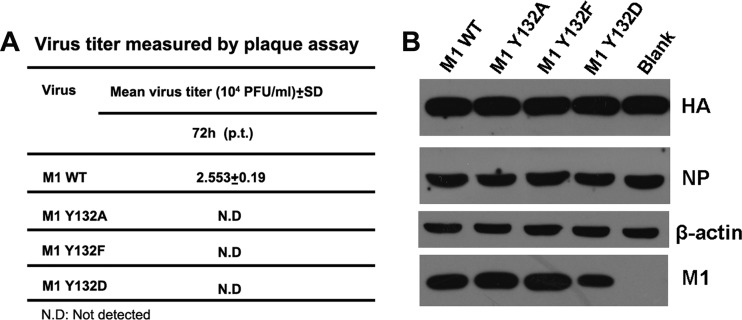
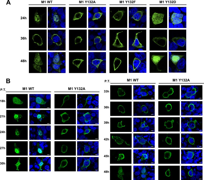
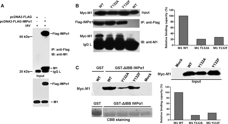
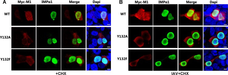
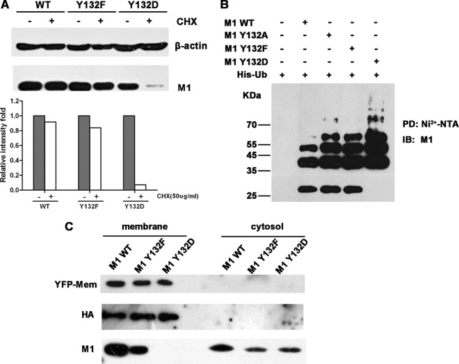
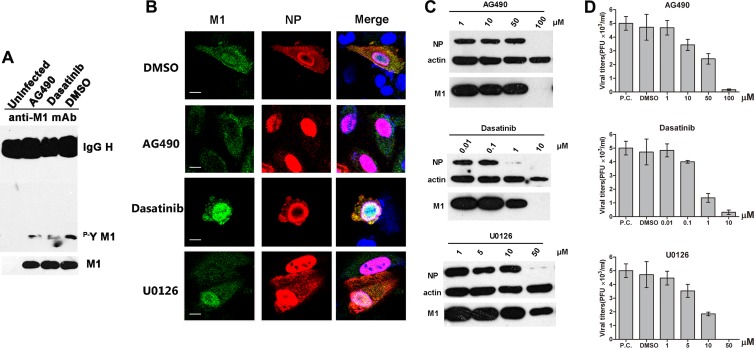
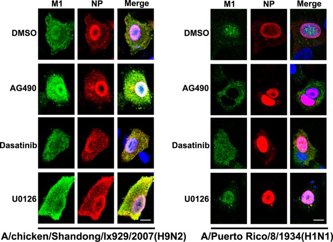
Phosphorylation of viral proteins plays important roles in the influenza A virus (IAV) life cycle. By using mass spectrometry, we identified tyrosine 132 (Y132) as a phosphorylation site of the matrix protein (M1) of the influenza virus A/WSN/1933(H1N1). Phosphorylation at this site is essential to the process of virus replication by controlling the nuclear import of M1. We further demonstrated that the phosphorylated tyrosine is crucial for the binding of M1 to the nuclear import factor importin-α1, since any substitutions at this site severely reduce this protein-protein interaction and damage the importin-α1-mediated nuclear import of M1. Additionally, the tyrosine phosphorylation which leads to the nuclear import of M1 is blocked by a Janus kinase inhibitor. The present study reveals a pivotal role of this tyrosine phosphorylation in the intracellular transportation of M1, which controls the process of viral replication.


Figures

Fig 1
Fig 1
Tyrosine 132 is a phosphorylation site on the influenza A virus M1 protein. (A) Gel electrophoresis of phosphorylated M1 protein immunoprecipitated with a monoclonal anti-M1 antibody. The bands were digested in the gel and then identified by LC-MS/MS analysis. The locations of M1 and phosphorylated M1 (pM1) are indicated by arrows. ALP, alkaline phosphatase. (B) The protein was identified as the influenza A virus matrix protein M1 [A/WSN/1933(H1N1)]. Peptides (blue) detected by MS and the phosphorylated tyrosine site (red) are indicated. (C) Mass spectrometry analysis was performed on the purified M1 protein. The fragment ions b18 and b19, with correspondingly increased phosphoric acid, indicate that Y132 is a modification site. (D) Surface accessibility of Y132 as analyzed using PyMOL. Y132 (yellow) and its functional hydroxyl group (red) are visible, which indicates that they are on the surface of the M1 protein. The NLS of M1 (101RKLKR105; blue) (PDB accession no. 1EA3) is also shown.
Fig 2
Fig 2
Tyrosine 132 is a key site of phosphotyrosine on M1. (A) Phosphotyrosine-containing M1 was detected by specific anti-p-Tyr antibodies in a virus rescue system, and 1/10 the amount of whole loading protein was probed with anti-M1 antibody to show the levels of total M1. The relative pY M1 intensity was determined as the ratio of tyrosine-phosphorylated M1 to total M1. The loss of phosphorylation at Y132 drastically reduced the level of total M1 tyrosine phosphorylation (lanes 3 and 4). (B) Y132 and the putative SH2 binding motif (YRNM) are greatly conserved in M1 proteins among different subtypes of influenza A virus. Sequence alignment of the specific M1 domain (yellow) was performed by MegAlign. The schematic diagram of M1 shows the functional domains involving Y132 (upper panel). WT, wild type; TID, transcription inhibition domain; NES, nuclear export signal; NLS, nuclear localization signal.
Fig 3
Fig 3
Phosphorylated Y132 of M1 is essential for virus rescue. A 12-plasmid reverse genetic system was used to rescue recombinant viruses with WT and mutated M1s (Y132A/F/D). (A) At 72 h p.t., the culture supernatants were harvested and subjected to plaque assays on MDCK cells. All recombinant viruses containing M1 mutants failed to be rescued. (B) At 72 h p.t., the transfected cells were lysed for Western blot analysis. HA, NP, and M1 were detected with respective antibodies. β-Actin was probed as a loading control. Blank, cells transfected without an added M1 plasmid.
Fig 4
Fig 4
Phosphorylated Y132 of M1 is essential for nuclear import of M1. (A) The localization of WT and mutated M1s (Y132A/F/D) was determined in a 12-plasmid reverse genetic system. At 24, 36, and 48 h p.t., transfected cells were fixed and detected by anti-M1 antibody (green). (B) M1s (WT and Y132A mutant) were detected by an anti-M1 monoclonal antibody (green) at different time points during virus recuse. Bars, 10 μm. The nucleus was stained with DAPI (4′,6-diamidino-2-phenylindole; blue).
Fig 5
Fig 5
Phosphorylated Y132 is crucial for binding of M1 to the nuclear import factor importin-α1. (A) The binding of M1 and importin-α1 (IMPα1) during virus infection was determined by coimmunoprecipitation. (B) Myc-M1 WT and Y132A and Y132F mutant proteins were coexpressed with FLAG-IMPα1 in 293T cells. FLAG-IMPα1 was immunoprecipitated with anti-FLAG agarose, and the associated Myc-M1 proteins were detected with the anti-M1 antibody. (C) Myc-M1 WT and Y132A and Y132F mutant proteins were expressed in 293T cells, and the cell lysates were incubated with GST-fused IMPα1 (ΔIBB [amino acids 60 to 529]). GST-ΔIBB IMPα1-bound M1s were blotted with the anti-M1 antibody. The relative binding capacities of M1s and IMPα1 were calculated by quantifying the ratio of IMPα1-bound Myc-M1s to input M1s. CBB, Coomassie brilliant blue; IAV, influenza A virus; IB, immunoblotting; IMP, importin; IgG L, IgG light chain; GST, glutathione S-transferase.
Fig 6
Fig 6
Phosphorylated Y132 is essential for the importin-α1-mediated nuclear import of M1. (A and B) The association of M1 and IMPα1 was determined by immunofluorescence assay. Myc-M1s and FLAG-IMPα1 were cotransfected into 293T cells and stained with anti-Myc (red) and anti-FLAG (green) antibodies, respectively. The nucleus was stained with DAPI (blue). Cells were incubated with (B) or without (A) IAV (multiplicity of infection [MOI] = 5) for 1 h and then treated with cycloheximide (CHX; 50 μg/ml) for 3 h before fixation. “Input” represents 1/10 the total M1 proteins in each binding reaction mix. Bars, 10 μm.
Fig 7
Fig 7
Y-to-D substitution at site 132 impairs the intracellular stability and membrane association of M1. (A) Twenty-four hours after transfection with constructs for WT, Y132F, or Y132D M1, 293T cells were treated with or without CHX (50 μg/ml) for 4 h. Cells were lysed and then detected with the anti-M1 antibody; β-actin was used as an internal control. The relative intensities were calculated by quantifying the results shown in the upper panel. (B) An in vivo ubiquitination assay suggested that mutation of Y to D increased the ubiquitination of the M1 protein. 293T cells were transfected with expression constructs for Myc-tagged M1s and His-tagged ubiquitin (His-Ub). At 40 hours posttransfection, the proteasome inhibitor MG-132 (10 μM) was added to the cells for 4 h. Cell extracts were then immunoprecipitated with Ni2+-NTA beads. The eluted proteins were analyzed by Western blotting using an anti-M1 antibody. PD, pulldown. (C) 293T cells were transfected with 12 plasmids from the virus rescue system and with an expression construct for a membrane marker, pEYFP-Mem (BD). After a 72-h transfection period, samples of cellular membrane and cytoplasmic fractions were separated and detected by anti-yellow fluorescent protein (anti-YFP), anti-HA, and anti-M1 antibodies.
Fig 8
Fig 8
The nuclear import of M1 is impaired by inhibiting Janus kinases. (A) A549 cells were infected with IAV (MOI = 0.1) for 12 h and then treated with the tyrosine kinase inhibitors AG490 (50 μM) and dasatinib (1 μM), with DMSO as a control (the inhibitors were dissolved in DMSO). After a 6-h treatment, cells were lysed and assayed for phosphotyrosine-containing M1. One-tenth the amount of whole loading protein was probed with anti-M1 antibody to show the levels of total M1. (B) A549 cells were infected with IAV (MOI = 2); after 1 h at 4°C for virus absorption, cells were washed with PBS and treated with several protein kinase inhibitors, including AG490 (100 μM), dasatinib (10 μM), and U0126 (50 μM), with DMSO as a control. Cells were fixed after 6 h of treatment and stained with anti-M1 (green) and anti-NP (red) antibodies. The nucleus was stained with DAPI (blue). Views of single cells with viral protein expression are shown. Bars, 10 μm. (C) A549 cells were infected with IAV (MOI = 2) for 1 h at 4°C, and then the medium was changed and cells were treated with kinase inhibitors at different concentrations. After 6 h of incubation at 37°C, cells were lysed and detected with anti-M1 and anti-NP antibodies. β-Actin was used as a loading control. (D) Virus titers of the supernatants in panel C were measured by plaque assay on MDCK cells. The positive control (P.C.) was IAV-infected cells without any additions (DMSO or inhibitors).
Fig 9
Fig 9
A Janus kinase inhibitor has different effects on nuclear localization of influenza A virus M1 proteins from different strains. A549 cells were infected with A/chicken/Shandong/lx929/2007(H9N2) or A/Puerto Rico/8/1934(H1N1) (MOI = 2); after 1 h at 4°C for virus absorption, cells were washed with PBS and then treated with AG490 (100 μM), dasatinib (10 μM), or U0126 (50 μM), with DMSO as a control. Cells were fixed after 6 h of treatment and then stained with anti-M1 (green) and anti-NP (red) antibodies. The nucleus was stained with DAPI (blue). Following staining, coverslips were analyzed using a model FV500 laser scanning confocal microscope. Views of single cells with viral protein expression are shown. Bars, 10 μm.

Similar articles

Cited by

References

    1. Samji T. 2009. Influenza A: understanding the viral life cycle. Yale J. Biol. Med. 82:153–159 - PMC - PubMed
    1. Watanabe T, Watanabe S, Kawaoka Y. 2010. Cellular networks involved in the influenza virus life cycle. Cell Host Microbe 7:427–439 - PMC - PubMed
    1. Jagger BW, Wise HM, Kash JC, Walters KA, Wills NM, Xiao YL, Dunfee RL, Schwartzman LM, Ozinsky A, Bell GL, Dalton RM, Lo A, Efstathiou S, Atkins JF, Firth AE, Taubenberger JK, Digard P. 2012. An overlapping protein-coding region in influenza A virus segment 3 modulates the host response. Science 337:199–204 - PMC - PubMed
    1. Wise HM, Hutchinson EC, Jagger BW, Stuart AD, Kang ZH, Robb N, Schwartzman LM, Kash JC, Fodor E, Firth AE, Gog JR, Taubenberger JK, Digard P. 2012. Identification of a novel splice variant form of the influenza A virus M2 ion channel with an antigenically distinct ectodomain. PLoS Pathog. 8:e1002998 doi:10.1371/journal.ppat.1002998 - DOI - PMC - PubMed
    1. Gregoriades A, Guzman GG, Paoletti E. 1990. The phosphorylation of the integral membrane (M1) protein of influenza virus. Virus Res. 16:27–41 - PubMed

Publication types

MeSH terms

LinkOut - more resources