. 2013 Jun;87(11):6246-56.
doi: 10.1128/JVI.03298-12. Epub 2013 Mar 27.

The receptor attachment function of measles virus hemagglutinin can be replaced with an autonomous protein that binds Her2/neu while maintaining its fusion-helper function

Affiliations

The receptor attachment function of measles virus hemagglutinin can be replaced with an autonomous protein that binds Her2/neu while maintaining its fusion-helper function

Anke Rasbach et al. J Virol. 2013 Jun.

Abstract

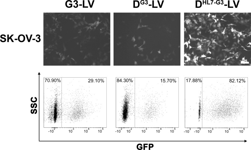
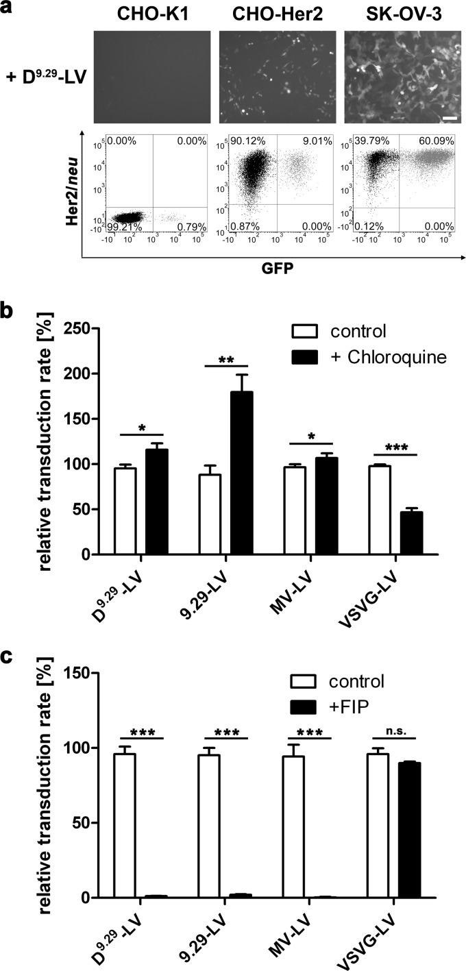
Cell entry of enveloped viruses is initiated by attachment to the virus receptor followed by fusion between the virus and host cell membranes. Measles virus (MV) attachment to its receptor is mediated by the hemagglutinin (H), which is thought to produce conformational changes in the membrane fusion protein (F) that trigger insertion of its fusion peptide into the target cell membrane. Here, we uncoupled receptor attachment and the fusion-helper function of H by introducing Y481A, R533A, S548L, and F549S mutations into the viral attachment protein that made it blind to its normal receptors. An artificial receptor attachment protein specific for Her2/neu was incorporated into the membranes of pseudotyped lentivirus particles as a separate transmembrane protein along with the F protein. Surprisingly, these particles entered efficiently into Her2/neu-positive SK-OV-3 as well as CHO-Her2 cells. Cell entry was independent of endocytosis but strictly dependent on the presence of H. H-specific monoclonal antibodies, as well as a mutation in H interfering with H/F cooperation, blocked cell entry. The particles mediated stable and specific transfer of reporter genes into Her2/neu-positive human tumor cells also in vivo, while exhibiting improved infectivity and higher titers than Her2/neu-targeted vectors displaying the targeting domain on H. Extending the current model of MV cell entry, the data suggest that receptor binding of H is not required for its fusion-helper function but that particle-cell contact in general may be sufficient to induce the conformational changes in the H/F complex and activate membrane fusion.


Figures

Fig 1
Fig 1
D9.29 colocalizes with MV glycoproteins at the cell surface of HEK-293T producer cells. (a) Schematic drawing of D9.29, FΔ30, HmutΔ18, and 9.29-HmutΔ18. DARPin 9.29 has been fused to the transmembrane domain (TMD) of the platelet-derived growth factor receptor (PDGFR) or the MV HmutΔ18 protein. Immunological tags (myc and HA) are indicated. Both MV envelope proteins are cytoplasmic tail (CT) truncated. HmutΔ18 carries four mutations (Y481A, R533A, S548L, and F549S) in the SLAM and CD46 recognition sites (19), which are indicated by asterisks. (b) HmutΔ18 and D9.29 coimmunostaining one day after HEK-293T cells were transfected with 0.25 μg of pCG-HmutΔ18, pCG-FΔ30, and pDisplay-D9.29. Fixed and permeabilized cells were stained with anti-H and then with anti-HA antibody. Secondary antibodies conjugated with Alexa Fluor dyes 546 and 633 were used for detection. Scale bar, 10 μm. (c) Surface expression of D9.29 on HEK-293T cells transiently transfected with pDisplay-D9.29 (black) compared to mock-transfected cells (gray) as determined by flow cytometry analysis. Cells were stained with PE-coupled anti-HA antibody. (d) Detergent-resistant membranes (DRM) from HEK-293T cells transfected with pCMVΔR8.9, pSEW, pCG-HmutΔ18, pCG-FΔ30, and pDisplay-D9.29 were separated by flotation assay. The distribution of proteins within fractions (fraction 1 represents the top of the gradient) and the pellet (P) was assessed by immunoblotting with antibodies specific for H, F, the HA tag of D9.29, the DRM marker flotillin 1, and the non-DRM marker CD46.
Fig 2
Fig 2
MV-pseudotyped virus incorporates D9.29. (a) Principle of virus particle generation by transfection of HEK-293T cells. Transfection with the plasmids shown gives rise to D9.29-LV particles pseudotyped with HmutΔ18, FΔ30, and D9.29. (b) Western blot analysis for incorporation of F1Δ30, D9.29, p24, and HmutΔ18 or 9.29-HmutΔ18 in vector particles 9.29-LV, D9.29-LV, and variants of D9.29-LV in which FΔ30 (w/o FΔ30), HmutΔ18 (w/o HmutΔ18), or D9.29 (w/o D9.29) was omitted, respectively, or HmutΔ18 was replaced by HmutΔ18* (w/HmutΔ18*). (c) Immunoelectron microscopy of ultrathin sections of HEK-293T producer cells releasing D9.29-LV vector particles. D9.29 was detected with primary antibody directed against the HA tag and secondary anti-mouse antibody conjugated with 10-nm gold particles following epoxy embedding and ultrathin sectioning. Scale bar, 200 nm.
Fig 3
Fig 3
D9.29-LV mediates efficient gene delivery. Equal amounts of egfp gene-delivering virus particles 9.29-LV, D9.29-LV, and variants of D9.29-LV in which FΔ30 (w/o FΔ30), HmutΔ18 (w/o HmutΔ18), or D9.29 (w/o D9.29) was omitted, respectively, or HmutΔ18 was replaced by H*mutΔ18 (w/H*mutΔ18) were added to SK-OV-3 cells. (a) Representative pictures and flow cytometry dot plots of SK-OV-3 cells 3 days after addition of virus. Scale bar, 100 μm. (b) Titers on SK-OV-3 cells as determined by flow cytometry analysis. Arrows indicate titers of <1 × 10⁴ transducing units (t.u.)/ml. (c) Infectivity determined as ratio of transducing units (t.u.) per μg p24 for 9.29-LV, D9.29-LV, and MV-LV (n = 4; mean ± standard deviations [SD] are shown; *, P < 0.01; n.s., not significant by unpaired t test).
Fig 4
Fig 4
High-affinity receptor binding is needed for virus entry. SK-OV-3 cells were incubated with equal amounts of egfp gene-delivering virus particles 9.29-LV, D9.29-LV, and variants of D9.29-LV in which FΔ30 (w/o FΔ30), HmutΔ18 (w/o HmutΔ18), or D9.29 (w/o D9.29) was omitted, respectively, or HmutΔ18 was replaced by H*mutΔ18 (w/H*mutΔ18) either without centrifugation (a) or with additional centrifugation at 700 × g for 1 h (b). Flow cytometry analysis was performed 72 h after transduction.
Fig 5
Fig 5
DARPin G3 as an alternative attachment protein. SK-OV-3 cells were transduced with particles presenting the Her2/neu-specific DARPin G3 fused to H (G3-LV), to the PDGFR transmembrane domain (DG3-LV), or to the PDGFR transmembrane domain connected by a helical linker (DHL7-G3-LV). Representative pictures of EGFP-expressing cells were taken at 72 h after transduction, and the percentage of EGFP-positive cells was determined by flow cytometry. Scale bar, 100 μm.
Fig 6
Fig 6
Characterization of D9.29-LV cell entry. (a) Cell entry is Her2/neu dependent. Representative pictures and flow cytometry dot plots of CHO-K1, CHO-Her2, and SK-OV-3 cells at 72 h after transduction with D9.29-LV are shown. Scale bar, 100 μm. (b) SK-OV-3 cells (1 × 10⁴) were treated with 150 μM chloroquine before the indicated virus particles were added. Relative transduction rates compared to that for cells transduced in the absence of chloroquine were determined by flow cytometry analysis. (c) MV-pseudotyped LV particles (0.07 μg p24) and VSVG-LV (0.008 μg p24) were incubated with (+FIP) or without (control) 200 μM FIP for 1 h at room temperature and then added to SK-OV-3 cells at an MOI of 0.3. The percentages of EGFP-positive cells were determined by flow cytometry after 72 h, and transduction rates relative to that obtained with untreated particles were calculated (n = 3; means ± SD are shown; *, P < 0.05; **, P < 0.01; ***, P < 0.0001; n.s., not significant by unpaired t test).
Fig 7
Fig 7
Neutralization of D9.29-LV by H-specific monoclonal antibodies. (a) Sphere representation of the H dimer crystal structure (58) (Protein Data Bank [PDB] ID 2ZB5; modified with PyMOL). The stalk, transmembrane, and cytoplasmic regions are depicted as vertical lines, with two horizontal lines indicating disulfide bonds. Putative antibody binding sites are indicated in orange, red, green, and blue. Residues mutated in HmutΔ18 to ablate natural receptor tropism are shown in yellow. Antigenic site vi is shown in purple. The virus membrane is illustrated as a gray box. (b) MV-pseudotyped LV (0.1 μg p24) and VSVG-LV (0.004 μg p24) particles were incubated with or without H-specific MAbs K71 (top left), L77 (top right), Nc32 (bottom left), and K29 (bottom right) for 1 h at room temperature and then added to SK-OV-3 cells at an MOI of 0.3. The percentages of EGFP-positive cells were determined by flow cytometry after 72 h, and transduction rates relative to that obtained in the absence of MAbs were calculated (n = 3; means ± SD are shown). (c) HEK-293T cells (1 × 10⁶) transiently transfected with pCG-HΔ18, pCG-HmutΔ18, or pCG-HmutΔ18-DARPin-9.29 were stained with K29, K71, Nc32, or L77. Secondary anti-mouse-PE was used for detection in flow cytometry. (d) Binding of virus particles to SK-OV-3 cells. D9.29-LV was incubated with 1 μg K29, K71, Nc32, or L77 for 1 h at room temperature, and then SK-OV-3 cells were added and left for another hour. Samples were stained against the HA tag of particles using an anti-HA PE-coupled antibody. Cells incubated only with virus or virus displaying an attachment domain unable to bind to Her2/neu (DX-LV) were used as positive and negative controls, respectively. Fluorescence of cell-particle complexes was analyzed using flow cytometry.
Fig 8
Fig 8
D9.29-LV as gene transfer vector. (a and b) Titers of D9.29-LV stocks were optimized by keeping the amount of pDisplay-D9.29 constant and varying that of pCG-FΔ30 to pCG-HmutΔ18 (a) or keeping the amounts of pCG-FΔ30 and pCG-HmutΔ18 constant and varying that of pDisplay-D9.29 (b) for transfection of HEK-293T cells along with pCMVΔ8.91 and pSEW. For each ratio, pseudotyped vectors were titrated on SK-OV-3 cells, and their relative titer normalized to that obtained after transfection of a 1:1 plasmid ratio (100%) is shown (n = 3; means ± SD are shown). (c) SK-OV-3 cells were transduced with D9.29-LV or VSVG-LV in the presence or absence of 10 μM reverse transcriptase inhibitor azidothymidine (AZT) to verify that vectors mediated transgene integration. Relative transduction efficiency is shown (n = 3; means ± SD are shown; P < 0.001 by unpaired t test). (d) In order to demonstrate stable transduction, SK-OV-3 cells were cultivated for 20 days after transduction with D9.29-LV at an MOI of 0.4 or 0.1. The percentage of EGFP-positive cells was determined at the indicated time points by flow cytometry analysis. (e) CB17 SCID mice were injected with 5 × 10⁶ SK-OV-3 cells into the right flank (arrowheads). Once tumors had reached a volume of about 50 mm3, D9.29-LVluc (8 μg p24) or MVmut-LVluc (15 μg p24) was injected into the tail vein, and luciferase signals were analyzed by in vivo imaging 1 week later. Luciferase signal intensity is expressed as photons/second/square centimeter/steradian.

Similar articles

Cited by

References

    1. Ader N, Brindley MA, Avila M, Origgi FC, Langedijk JPM, CÖrvell Vandevelde M, Zurbriggen A, Plemper RK, Plattet P. 2012. Structural rearrangements of the central region of the morbillivirus attachment protein stalk domain trigger F protein refolding for membrane fusion. J. Biol. Chem. 287:16324–16334 - PMC - PubMed
    1. Apte-Sengupta S, Negi S, Leonard VHJ, Oezguen N, Navaratnarajah CK, Braun W, Cattaneo R. 2012. Base of the measles virus fusion trimer head receives the signal that triggers membrane fusion. J. Biol. Chem. 287:33026–33035 - PMC - PubMed
    1. Paal T, Brindley MA, St Clair C, Prussia A, Gaus D, Krumm SA, Snyder JP, Plemper RK. 2009. Probing the spatial organization of measles virus fusion complexes. J. Virol. 83:10480–10493 - PMC - PubMed
    1. Plemper RK, Hammond AL, Cattaneo R. 2001. Measles virus envelope glycoproteins hetero-oligomerize in the endoplasmic reticulum. J. Biol. Chem. 276:44239–44246 - PubMed
    1. Iorio RM, Melanson VR, Mahon PJ. 2009. Glycoprotein interactions in paramyxovirus fusion. Future Virol. 4:335–351 - PMC - PubMed

Publication types