. 2013 Jun;87(11):6377-90.
doi: 10.1128/JVI.03557-12. Epub 2013 Mar 27.

Sigma-1 receptor regulates early steps of viral RNA replication at the onset of hepatitis C virus infection

Affiliations

Sigma-1 receptor regulates early steps of viral RNA replication at the onset of hepatitis C virus infection

Martina Friesland et al. J Virol. 2013 Jun.

Abstract

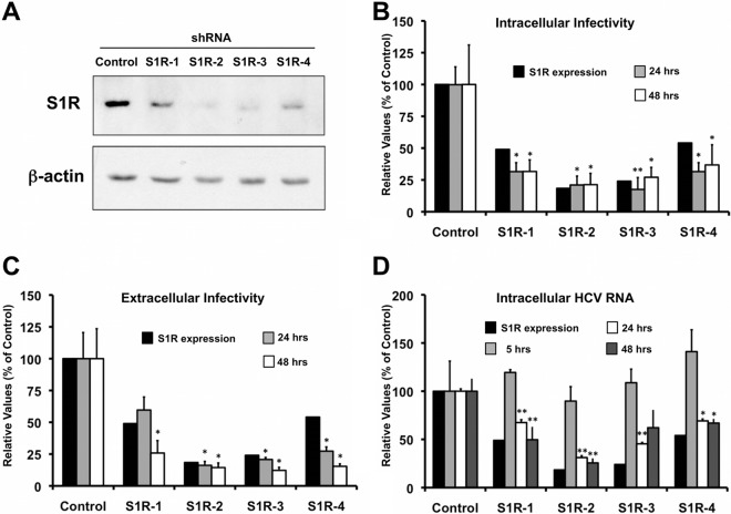
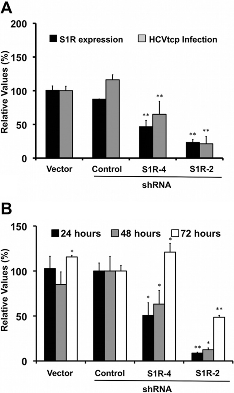
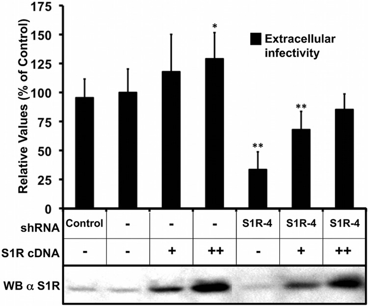
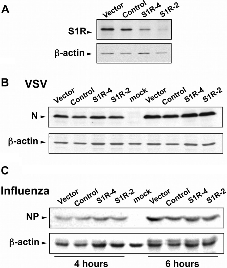
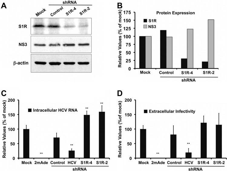
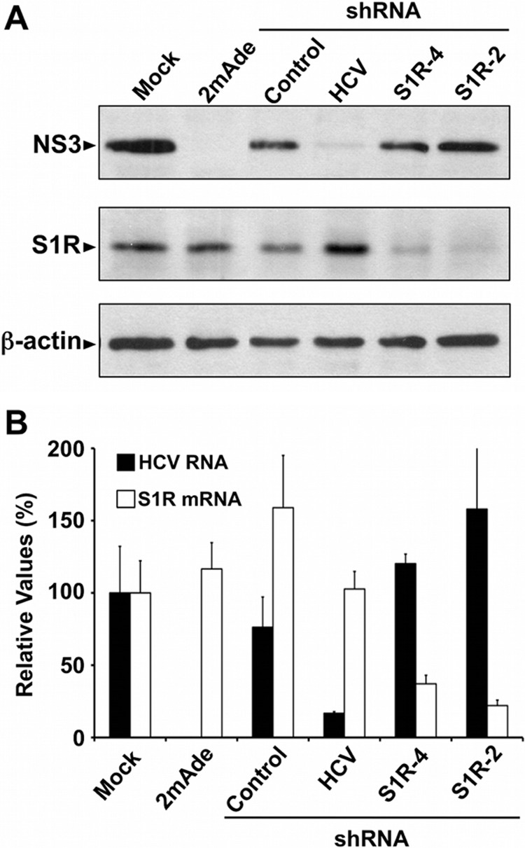
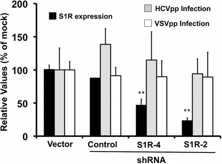
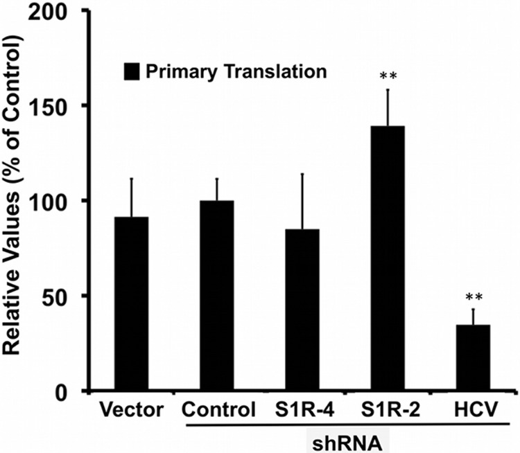
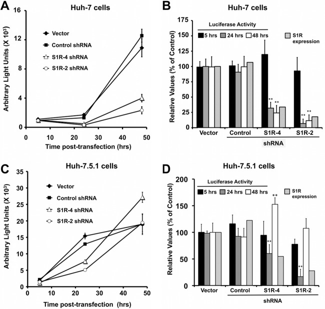
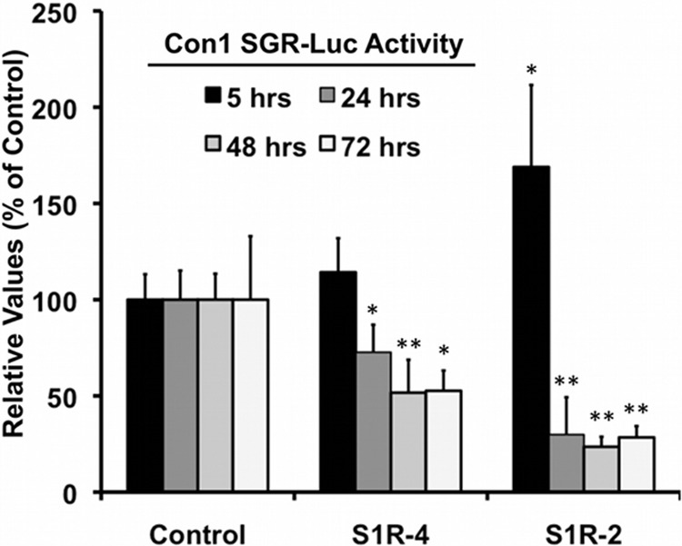
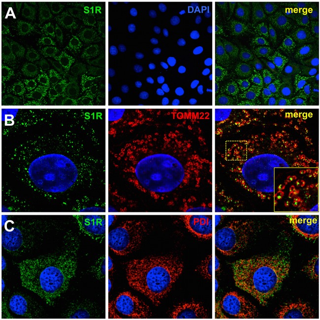
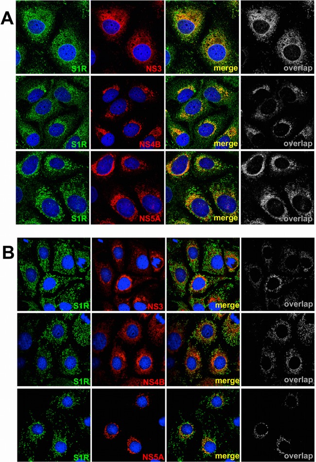
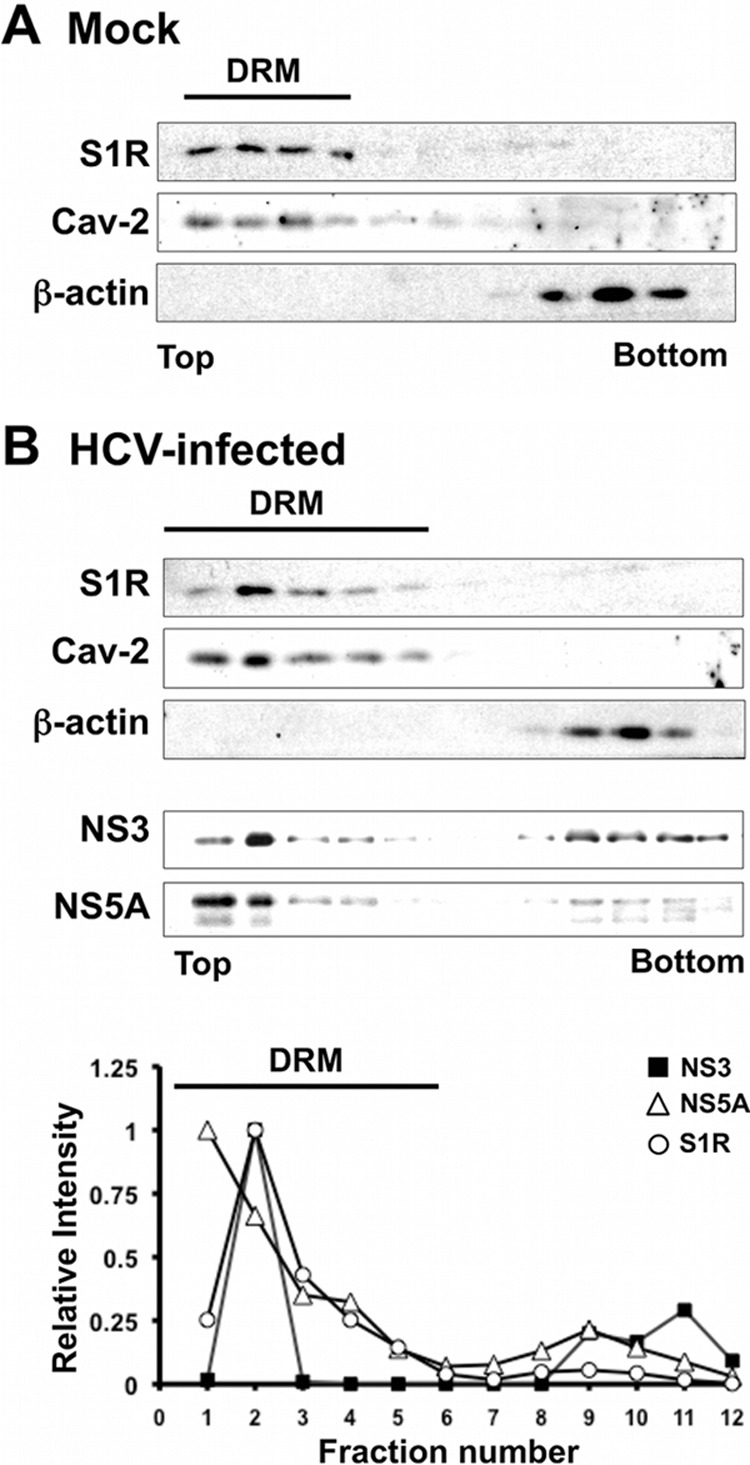
Hepatitis C virus (HCV) genome replication is thought to occur in a membranous cellular compartment derived from the endoplasmic reticulum (ER). The molecular mechanisms by which these membrane-associated replication complexes are formed during HCV infection are only starting to be unraveled, and both viral and cellular factors contribute to their formation. In this study, we describe the discovery of nonopioid sigma-1 receptor (S1R) as a cellular factor that mediates the early steps of viral RNA replication. S1R is a cholesterol-binding protein that resides in lipid-rich areas of the ER and in mitochondrion-associated ER membranes (MAMs). Several functions have been ascribed to this ER-resident chaperone, many of which are related to Ca(2+) signaling at the MAMs and lipid storage and trafficking. Downregulation of S1R expression by RNA interference (RNAi) in Huh-7 cells leads to a proportional decrease in susceptibility to HCV infection, as shown by reduced HCV RNA accumulation and intra- and extracellular infectivity in single-cycle infection experiments. Similar RNAi studies in persistently infected cells indicate that S1R expression is not rate limiting for persistent HCV RNA replication, as marked reduction in S1R in these cells does not lead to any decrease in HCV RNA or viral protein expression. However, subgenomic replicon transfection experiments indicate that S1R expression is rate limiting for HCV RNA replication without impairing primary translation. Overall, our data indicate that the initial steps of HCV infection are regulated by S1R, a key component of MAMs, suggesting that these structures could serve as platforms for initial RNA replication during HCV infection.


Figures

Fig 1
Fig 1
Cellular levels of S1R are limiting for HCV infection. Huh-7 cells were transduced with lentiviral vectors expressing an irrelevant sequence or shRNAs targeting S1R mRNA and infected at an MOI of 10 with D183v. (A) Western blot analysis performed on total cell extracts showing reduced S1R expression in shRNA-expressing cell lines at the time of inoculation (6 days posttransduction) and a loading control (β-actin). (B and C) Intracellular (B) and extracellular (C) infectivity titers (FFU/ml) in HCV-infected cells expressed as averages and standard deviations (n = 3). (D) Normalized HCV RNA levels were determined at 5, 24, and 48 h postinfection by RT-qPCR as described in Materials and Methods and are expressed as averages and standard deviations (n = 3). Normalized S1R expression in panels B and C was quantified from panel A. The statistical significance of the differences with the control data set was determined using Student's t test (*, P < 0.05; **, P < 0.01).
Fig 2
Fig 2
Cellular levels of S1R are limiting for HCVtcp infection. Huh-7 cells were transduced with lentiviral vectors expressing irrelevant sequences or shRNAs targeting S1R mRNA and infected with HCVtcp. (A) Twenty-four hours postinfection, cell lysates were used to measure luciferase activity and S1R expression. The data are shown as average percentages of the empty vector and standard deviations (n = 9). (B) Extended kinetics of HCVtcp infection at 24, 48, and 72 h postinfection. The data are shown as average percentages of the control vector and standard deviations for a representative experiment (n = 3). The statistical significance of the differences with the control data set was determined using Student's t test (*, P < 0.05; **, P < 0.01).
Fig 3
Fig 3
Expression of S1R cDNA restores susceptibility to infection of S1R-deficient cells. Mock-treated Huh7 cells and S1R shRNA4-expressing cells were transduced with a lentiviral vector expressing an shRNA-resistant S1R cDNA. The cells were subsequently infected at an MOI of 10 with D183v. Extracellular infectivity titers were determined at 24 h postinfection. The data are shown as averages and standard deviations of two independent experiments performed in triplicate (n = 6) and are represented as percentages of the mock-treated cells. The statistical significance of the differences with the control data set was determined using Student's t test (*, P < 0.05; **, P < 0.01). WB, Western blot; α S1R, anti-S1R; +, transduction with lentiviral vector; ++, transduction with higher dose of vector; −, no transduction.
Fig 4
Fig 4
S1R is not rate limiting for other RNA virus infections. Huh-7 cells were transduced with lentiviral vectors expressing irrelevant sequences or shRNAs targeting S1R mRNA. (A) Western blot analysis against S1R and a loading control (β-actin). (B and C) The cells were subsequently infected (MOI = 10) with VSV (B) or influenza virus (C). Protein samples were collected at 4 and 6 h postinfection and analyzed by Western blotting against VSV N (B) or influenza virus NP (C).
Fig 5
Fig 5
S1R downregulation does not interfere with persistent HCV infection. Persistently infected Huh-7 cells were untreated (Mock) or transduced with lentiviral vectors expressing an irrelevant shRNA (Control), an HCV-targeting shRNA (shRNA-HCV), or S1R-targeting (S1R-4 and S1R-2) shRNAs. Parallel cultures were treated with a specific HCV polymerase inhibitor (2mAde; 10 μM). Samples of the cells and supernatants were collected at day 9 posttransduction. (A) Western blotting against HCV NS3 protein (NS3), S1R, and loading control (β-actin) from one representative experiment of two. (B) Quantitation of S1R and NS3 protein expression normalized to the loading control. (C) Normalized intracellular HCV RNA levels determined by RT-qPCR. (D) Extracellular HCV infectivity titers. The data in panels C and D are shown as averages and standard deviations for two independent experiments performed in triplicate (n = 6) and are represented as percentages of the mock-treated cells. The statistical significance of the differences with the control data set was determined using Student's t test (**, P < 0.01).
Fig 6
Fig 6
S1R downregulation does not interfere with steady-state subgenomic JFH-1 replication. Huh-7 cells bearing a subgenomic JFH-1 replicon were transduced with an empty vector or with lentiviral vectors expressing an irrelevant shRNA (Control), an HCV-targeting shRNA (HCV), or S1R-targeting (S1R-4 and S1R-2) shRNAs. Parallel cultures were treated with a specific HCV polymerase inhibitor (2mAde; 10 μM). Samples of the cells were collected at day 6 posttransduction. (A) Western blotting against HCV NS3 protein (NS3), S1R, and loading control (β-actin). (B) Normalized intracellular HCV RNA and S1R mRNA levels determined by RT-qPCR. The data are shown as averages and standard deviations of triplicate (n = 3) samples from a representative experiment and are represented as percentages of the cells treated with an empty vector.
Fig 7
Fig 7
HCV entry is not altered by S1R downregulation. Huh-7 cells were transduced with lentiviral vectors expressing an empty vector (Vector), an irrelevant shRNA (Control) or S1R-targeting (shRNA4 and shRNA2) shRNAs. Once downregulation was verified by Western blotting (black bars), the cells were inoculated with pseudotyped retroviruses bearing JFH-1 (HCVpp) or VSV (VSVpp) envelope glycoproteins. Luciferase activity levels were determined at 48 h in total cell extracts. Normalized S1R expression levels were determined by Western blotting against S1R and β-actin. The data are shown as averages and standard deviations (n = 12) for four independent experiments performed in triplicate and are represented as percentages of cells transduced with an empty vector. The statistical significance of the differences with the control data set was determined using Student's t test (**, P < 0.01).
Fig 8
Fig 8
S1R expression levels are not limiting for primary translation. Huh-7 cells were transduced with lentiviral vectors expressing an irrelevant shRNA (Control), an HCV-targeting shRNA (HCV), or S1R-targeting (S1R-4 and S1R-2) shRNAs. S1R-deficient and control cells were transfected with a subgenomic JFH-1 replicon bearing a luciferase reporter gene and a polymerase activity-inactivating (GND) mutation. Luciferase activity was measured 5 h posttransfection. The data are shown as averages and standard deviations (n = 6) for two independent experiments performed in triplicate and are represented as percentages of cells transduced with the control vector. The statistical significance of the differences with the control data set was determined using Student's t test (**, P < 0.01).
Fig 9
Fig 9
S1R expression levels are limiting for initiation of HCV RNA replication. S1R-deficient cells were transfected with a subgenomic JFH-1 replicon bearing a luciferase reporter gene. Luciferase activity was measured at the indicated time points. S1R expression was quantitated by Western blotting. (A) Representative transfection experiment. Data are shown as averages and standard deviations (n = 3) of the measured light units. (B) Averages and standard deviations for three independent transfection experiments performed in triplicate (n = 9) represented as percentages of the cells transduced with an empty vector. (C and D) Similar experiments were conducted in Huh-7.5.1. cells (n = 6). The statistical significance of the differences with the control data set was determined using Student's t test (**, P < 0.01).
Fig 10
Fig 10
S1R expression is rate limiting for early genotype 1b HCV RNA replication. Control and S1R-deficient Huh-7 cells were prepared by lentiviral transduction of Huh-7 cells. S1R-deficient and control cells were electroporated with an in vitro-transcribed dicistronic HCV subgenomic replicon RNA from genotype 1b (Con1) bearing a luciferase reporter gene. Samples were collected at the indicated time points, and relative replication was determined as described in Materials and Methods. The data are shown as averages and standard deviations for two independent experiments performed in triplicate (n = 6). The statistical significance of the differences with the control data set was determined using Student's t test (*, P < 0.05; **, P < 0.01).
Fig 11
Fig 11
S1R is associated with MAMs. (A) Confocal immunofluorescence microscopy of S1R in Huh-7 cells displaying two different subcellular patterns. (B and C) Higher-magnification images showing S1R (green) colocalization with mitochondria (TOMM22; red) in discrete cytoplasmic punctae (B) (the boxed area in the merged image is shown enlarged in the inset) and with ER markers (PDI; red) in a diffuse cytoplasmic staining (C). Nuclei were stained with DAPI and are shown in blue.
Fig 12
Fig 12
S1R colocalizes with HCV NS proteins. Confocal immunofluorescence microscopy of HCV-infected Huh-7 cells (MOI = 10) at 48 (A) and 72 (B) h postinfection. S1R is shown in green, and NS3, NS4B, and NS5A are shown in red. Representative images from three independent experiments are shown. Colocalization masks (overlap) are shown in grayscale.
Fig 13
Fig 13
S1R cofractionates with HCV NS proteins in detergent-resistant membranes. Cell lysates were produced by incubating HCV-infected Huh-7 cells in TNE-0.5% Triton X-114. The cleared lysates were placed onto a discontinuous 10 to 40% sucrose-TNE gradient and centrifuged at 120,000 × g for 16 h. Gradient fractions were collected from the top and analyzed by Western blotting for the presence of NS3, NS5A, S1R, β-actin, and caveolin-2. (A) Western blot analysis of gradient fractions in mock-infected cells. (B) (Top) Western blot analysis of the gradient fractions in HCV-infected cells. (Bottom) Quantitation of S1R, NS3, and NS5A in the top blot. The values are shown as relative abundance per fraction relative to the maximum signal. Detergent-resistant membrane fractions containing caveolin-2 are marked as DRM.

References

    1. Guidotti LG, Chisari FV. 2006. Immunobiology and pathogenesis of viral hepatitis. Annu. Rev. Pathol. 1:23–61 - PubMed
    1. Poordad F, McCone J, Jr, Bacon BR, Bruno S, Manns MP, Sulkowski MS, Jacobson IM, Reddy KR, Goodman ZD, Boparai N, DiNubile MJ, Sniukiene V, Brass CA, Albrecht JK, Bronowicki JP. 2011. Boceprevir for untreated chronic HCV genotype 1 infection. N. Engl. J. Med. 364:1195–1206 - PMC - PubMed
    1. Jacobson IM, McHutchison JG, Dusheiko G, Di Bisceglie AM, Reddy KR, Bzowej NH, Marcellin P, Muir AJ, Ferenci P, Flisiak R, George J, Rizzetto M, Shouval D, Sola R, Terg RA, Yoshida EM, Adda N, Bengtsson L, Sankoh AJ, Kieffer TL, George S, Kauffman RS, Zeuzem S. 2011. Telaprevir for previously untreated chronic hepatitis C virus infection. N. Engl. J. Med. 364:2405–2416 - PubMed
    1. Otto GA, Puglisi JD. 2004. The pathway of HCV IRES-mediated translation initiation. Cell 119:369–380 - PubMed
    1. Moradpour D, Penin F, Rice CM. 2007. Replication of hepatitis C virus. Nat. Rev. Microbiol. 5:453–463 - PubMed

Publication types