. 2013 May;24(10):1519-28.
doi: 10.1091/mbc.E12-11-0796. Epub 2013 Mar 27.

Ndufaf5 deficiency in the Dictyostelium model: new roles in autophagy and development

Affiliations

Ndufaf5 deficiency in the Dictyostelium model: new roles in autophagy and development

Sergio Carilla-Latorre et al. Mol Biol Cell. 2013 May.

Abstract

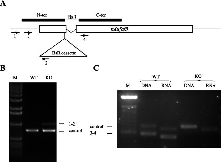
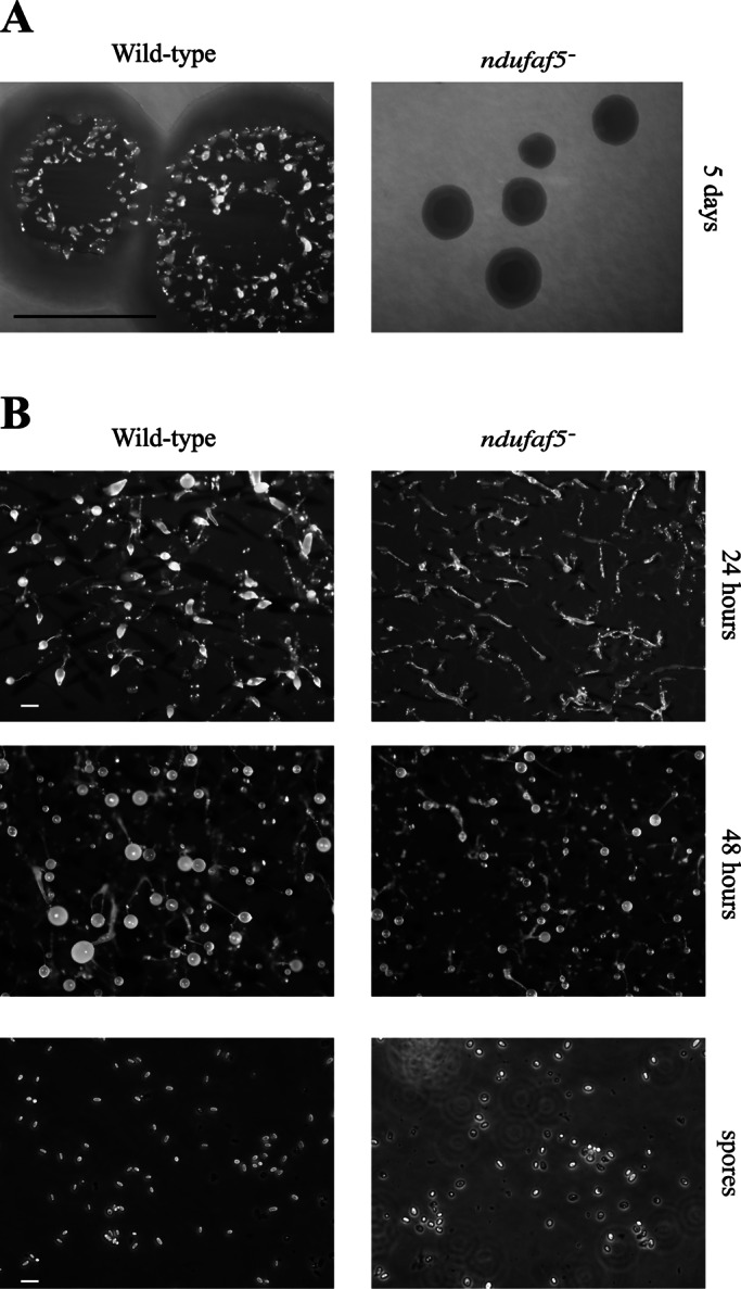
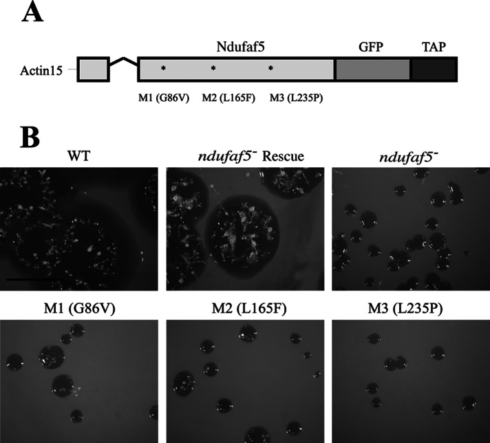
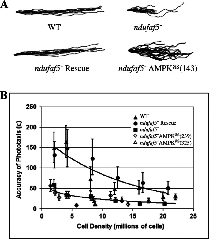
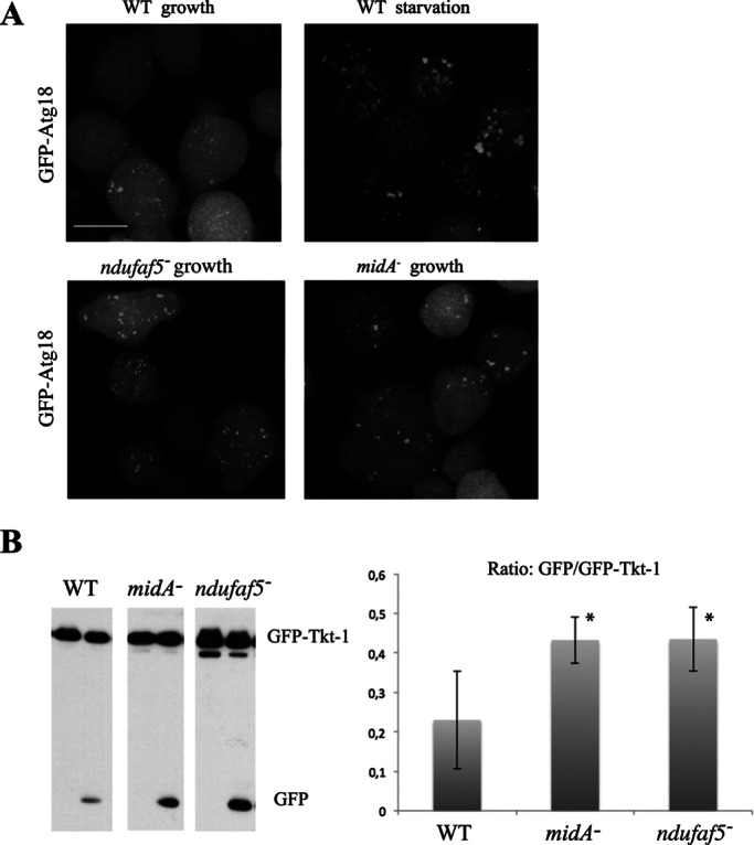
Ndufaf5 (also known as C20orf7) is a mitochondrial complex I (CI) assembly factor whose mutations lead to human mitochondrial disease. Little is known about the function of the protein and the cytopathological consequences of the mutations. Disruption of Dictyostelium Ndufaf5 leads to CI deficiency and defects in growth and development. The predicted sequence of Ndufaf5 contains a putative methyltransferase domain. Site-directed mutagenesis indicates that the methyltransferase motif is essential for its function. Pathological mutations were recreated in the Dictyostelium protein and expressed in the mutant background. These proteins were unable to complement the phenotypes, which further validates Dictyostelium as a model of the disease. Chronic activation of AMP-activated protein kinase (AMPK) has been proposed to play a role in Dictyostelium and human cytopathology in mitochondrial diseases. However, inhibition of the expression of AMPK gene in the Ndufaf5-null mutant does not rescue the phenotypes associated with the lack of Ndufaf5, suggesting that novel AMPK-independent pathways are responsible for Ndufaf5 cytopathology. Of interest, the Ndufaf5-deficient strain shows an increase in autophagy. This phenomenon was also observed in a Dictyostelium mutant lacking MidA (C2orf56/PRO1853/Ndufaf7), another CI assembly factor, suggesting that autophagy activation might be a common feature in mitochondrial CI dysfunction.


Figures

FIGURE 1:
FIGURE 1:
ndufaf5 disruption and loss of expression. (A) Scheme of the disruption vector and the disrupted gene after homologous recombination. The BsR cassette interrupted the gene at the nucleotide 206 of the coding sequence and deleted the intron (indicated as a V-shaped line). Open boxes represent ndufaf5 exons. Solid boxes represent the disruption vector with the position of the N and C-terminal fragments used to allow homologous recombination. The BsR cassette is located between the fragments and is not depicted in scale to allow the correct overlapping with the scheme of ndufaf5 locus. The arrowheads with numbers indicate the different oligonucleotides used for verification of the disruptant strains (Supplemental Table S1). (B) Genomic verification. DNA extracted from wild-type and knockout strains was subjected to PCR using oligos 1 and 2. The amplification band indicates that homologue recombination has taken place in the expected region. The unrelated gene (dictyBase ID: DDB_G0268840) was used as a PCR control. (C) Transcriptomic verification. RNA extracted from wild-type and knockout strains at 14 h of development was subjected to RT-PCR using oligos 3 and 4. The absence of amplification in the mutant strains indicates that the transcript has been disrupted. Differences in the size of the bands between DNA and RNA templates are due to the presence of a small intron in both ndufaf5 and the control (DDB_G0268840) genes. M, DNA marker.
FIGURE 2:
FIGURE 2:
Analysis of growth and development in ndufaf5. (A) Wild-type and ndufaf5 cells were seeded on SM plates in association with K. aerogenes and grown for 5 d. The ndufaf5 strain showed a strong defect in the diameter of the clearing zone. Bar, 1 cm. (B) Cells grown exponentially in axenic medium were harvested, washed with PDF buffer, and deposited onto nitrocellulose filters according to Materials and Methods. Photographs were taken at the indicated times. The mutant strain showed a delay at the finger-slug stage. Structures culminated after 48 h, and the fruiting bodies were smaller than those of wild type. Bar, 2 mm. Wild-type and mutant spores were taken from 10-d-old fruiting bodies and observed under phase contrast microscopy. Mutant spores were rounder and less refractive, suggesting loss of viability. Bar, 10 μm.
FIGURE 3:
FIGURE 3:
Functional conservation of Ndufaf5 and mitochondrial phenotypes in the ndufaf5 strain. (A) Ndufaf5-GFP was expressed in the mutant background and subcellular localization of the protein analyzed by confocal microscopy. The region marked was magnified below and the color intensity adjusted to allow a clearer visualization of mitochondria. Bar, 10 μm. (B) Activities of complexes I, II, III, and IV in ndufaf5 and wild-type cells were measured by spectrophotometric analyses and corrected by protein and citrate synthase activity and finally normalized against wild-type levels. (C) Citrate synthase and cellular ATP levels. Citrate synthase activity was measured by spectrophotometric analyses and normalized as before. ATP levels were measured using a bioluminescence assay and normalized vs. wild-type levels. NS, nonsignificant. **p < 0.01.
FIGURE 4:
FIGURE 4:
Site-directed mutagenesis analyses. (A) Schematic representation of the constructs used for complementation or site-directed mutagenesis experiments. Ndufaf5 was fused to GFP and tandem affinity purification tag (TAP-tag) into the pDV-GFP-CTAP vector, where expression is directed under the control of actin-15 promoter. The V-shaped line represents an intron. M1 is the mutation in the predicted SAM-binding domain (see the text). M2 and M3 are the corresponding mutations to the pathogenic ones in human (M2 equivalent to L159F, and M3 equivalent to L229P). (B) Wild-type and mutant constructs were transfected in ndufaf5 cells. The expressed proteins colocalized with MitoTracker Red in mitochondria to the same extent as depicted in Figure 3A (data not shown). Transfected cells were spread onto SM plates to test the size of the clearing zone after 5 d. The strain transformed with the wild-type protein (ndufaf5 rescue strain) complemented the growth phenotype completely, in contrast to the mutated forms, which showed similar defects as the parental ndufaf5. Bar, 1 cm.
FIGURE 5:
FIGURE 5:
Phototaxis defects in ndufaf5 are not rescued by AMPK inhibition. (A) Qualitative experiments. Different colonies (slugs) were allowed to migrate under lateral light source. Pictures were obtained after 48 h of incubation at 21ºC and the slug trails represented (see Materials and Methods). The ndufaf5 slugs had a significant defect in phototaxis, which was rescued after transfection with the Ndufaf5 wild-type protein (ndufaf5 rescue). However, no suppression of the mutant phenotype was observed in ndufaf5 cells expressing a different number of copies of an AMPK antisense construct. A representative AMPK-inhibited strain is shown, with the number of copies between brackets. (B) Quantitative experiments. Phototaxis accuracy (κ value) was calculated for all the indicated strains at different cell densities (see Materials and Methods). A clear defect was again observed in ndufaf5 migrating slugs. This defect was complemented by overexpression of Ndufaf5 (ndufaf5 rescue) but not suppressed by antisense inhibition of AMPK (number of copies of AMPKas construct in brackets). The error bars represent 90% confidence intervals. Lines of best fit were determined by the least squares method, fitting an exponential model (wild type and ndufaf5 rescue) or logarithmic model (ndufaf5 mutant and AMPK antisense strains derived from it). The choice of model was dictated by which provided the best fit by eye.
FIGURE 6:
FIGURE 6:
Autophagy is increased in ndufaf5 and midA cells. (A) Labeling of autophagosomes. The indicated strains were transfected with the autophagosome marker GFP-Atg18. Cells were incubated in growth media or starvation conditions (PDF buffer). Representative images show maximum projections of confocal images covering the whole cell. Bar, 10 μm. (B) Autophagic flux. Strains transfected with the marker GFP-Tkt-1 growing in HL5 were incubated with 0 or 100 mM NH4Cl (left and right lanes, respectively) as described in Materials and Methods. Extracts containing the same protein load were subjected to SDS–PAGE, transferred by Western blot, and incubated with α-GFP antibody to detect GFP-Tkt-1 and the cleaved GFP proteins. Left, a representative image of Western blots. Right, quantification of the ratio between free GFP and GFP–Tkt-1 of three independent experiments. *p < 0.05.

References

    1. Adachi H, Hasebe T, Yoshinaga K, Ohta T, Sutoh K. Isolation of Dictyostelium discoideum cytokinesis mutants by restriction enzyme-mediated integration of the blasticidin S resistance marker. Biochem Biophys Res Commun. 1994;205:1808–1814. - PubMed
    1. Annesley SJ, Fisher PR. Dictyostelium discoideum—a model for many reasons. Mol Cell Biochem. 2009a;329:73–91. - PubMed
    1. Annesley SJ, Fisher PR. Dictyostelium slug phototaxis. Methods Mol Biol. 2009b;571:67–76. - PubMed
    1. Bokko PB, Francione L, Bandala-Sanchez E, Ahmed AU, Annesley SJ, Huang X, Khurana T, Kimmel AR, Fisher PR. Diverse cytopathologies in mitochondrial disease are caused by AMP-activated protein kinase signaling. Mol Biol Cell. 2007;18:1874–1886. - PMC - PubMed
    1. Bridges HR, Fearnley IM, Hirst J. The subunit composition of mitochondrial NADH:ubiquinone oxidoreductase (complex I) from Pichia pastoris. Mol Cell Proteomics. 2010;9:2318–2326. - PMC - PubMed

Publication types