. 2013;8(3):e60543.
doi: 10.1371/journal.pone.0060543. Epub 2013 Mar 25.

Characterization and functional analysis of the potato pollen-specific microtubule-associated protein SBgLR in tobacco

Affiliations

Characterization and functional analysis of the potato pollen-specific microtubule-associated protein SBgLR in tobacco

Chen Liu et al. PLoS One. 2013.

Abstract

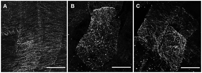
Microtubule-associated proteins play a crucial role in the regulation of microtubule dynamics, and are very important for plant cell and organ development. SBgLR is a potato pollen-specific protein, with five imperfect V-V-E-K-K-N/E-E repetitive motifs that are responsible for microtubule binding activity. In present study, SBgLR showed typical microtubule-associated protein characteristics; it bound tubulin and microtubules, and colocalized with microtubules in vitro. We also found that SBgLR could form oligomers, and that both the SBgLR monomers and oligomers bundle microtubules in vitro. Constitutive expression of SBgLR in tobacco caused curving and right-handed twisting root growth, abnormal directional cell expansion and cell layer arrangement, and pollen abortion. Immunofluorescence staining assays revealed that microtubule organization is altered in root epidermal cells in SBgLR-overexpressing lines. These suggest that SBgLR functions as a microtubule-associated protein in pollen development. Our results indicate that normal organization of MTs may be crucial for pollen development.


Conflict of interest statement

Competing Interests: The authors have declared that no competing interests exist.

Figures

Figure 1
Figure 1. Gene structure of SBgLR and amino acid comparison of SBgLR and its homologs.
A, Gene structure of SBgLR. Three exons were illustrated in black. B, Amino acid comparison between SBgLR and its homologs: St901, TSB, MAP18 and SB401. The conserved motifs that responsible for MT-binding domain were shadowed in gray.
Figure 2
Figure 2. Coomassie brilliant blue stained gels of recombinant SBgLR protein and tubulin.
A, Lane 1, 15 µg of total extract from bacteria cells containing pET30a empty vector without IPTG induction; Lane 2, 15 µg of total extract from bacterial cells containing pET30a-SBgLR vector without induction; Lane 3, 20 µg of total extract from bacteria cells containing pET30a-SBgLR vector with 50 mM IPTG induction for 4 h; Lane 4, 10 µg purified SBgLR recombinant protein. B, Lane 1, crude extract of porcine tubulin; Lane 2, purified porcine tubulin. The sizes of protein marker were labeled in the ‘kD’ column.
Figure 3
Figure 3. The recombinant SBgLR binds tubulin and MTs in vitro.
A to D, Immunoblotting assay. Aliquots of 2, 4, 8, and 16 µM of SBgLR, tubulin, and BSA were spotted onto PVDF membranes. The membrane was preincubated with (B) or without (A) 10 µM recombinant SBgLR, and then probed with anti-SBgLR antibody. The membrane was preincubated with (D) or without (C) 10 µM tubulin, and then probed with anti-β-tubulin antibody. E and F, The co-sedimentation assay. E, Coomassie brilliant blue stained gels of the pellets (P) and the supernatant (S) from the co-sedimentation assay. The recombinant SBgLR protein mainly appeared in the supernatant (S) after the centrifugation in the absence of tsMTs and co-precipitated with tsMTs into the pellets (P) when tsMTs was added. BSA (12 µM) was used as a negative control. F, Quantitative analysis of the binding between recombinant SBgLR and tsMTs. The binding of the recombinant SBgLR protein to MTs was saturated at a ratio of ∼0.35 mole recombinant SBgLR per mole tubulin dimer estimated by gel scanning.
Figure 4
Figure 4. SBgLR protein bundles MTs in vitro.
A to C, Confocal microscopy of recombinant SBgLR on MT-bundling. A, tsrMTs. Single-filament tsrMTs were scattered throughout the solution, no MT-bundles were observed. B, MT-bundles were formed when recombinant SBgLR was added. C, boiling denatured recombinant SBgLR was added as a negative control. Bar = 25 µm from A to C. D and E, Negative-staining electron microscopy of recombinant SBgLR on MT-bundling. D, tsMTs. Bar = 1 µm. E, Scattered tsMTs formed aggregates and integrated into a meshwork (black arrows) when native recombinant SBgLR was added. Bar = 100 nm. F, Longitudinal section of the MT-bundles. The cross-bridges were observed between the tsMTs (white arrows). Bar = 200 nm. G to L, Co-localization assay using confocal microscopy. G to I, tsrMTs were incubated with recombinant SBgLR, and then incubated with anti-SBgLR primary antibody and FITC conjugated goat anti-rabbit IgG secondary antibody. G, tsrMTs; H, recombinant SBgLR; I, Merged image of G and H. J to L, tsrMTs were incubated with boiling denatured recombinant SBgLR as a negative control. No MT-bundles and SBgLR signals were detected. J, tsrMTs; K, SBgLR; L, merged image of J and K. Bar = 10 µm from G to L. M to O, Confocal microscopy of recombinant SBgLR monomers on MT-bundling. M, tsrMTs. N, MT-bundles were formed when β-mercaptoethanol-treated recombinant SBgLR was added to tsrMTs. O, boiling denatured β-mercaptoethanol-treated recombinant SBgLR was added to tsrMTs as a negative control. Bar = 25 µm from M to O. P to R, Negative-staining electron microscopy of β-mercaptoethanol-treated recombinant SBgLR on MTs-bundling. P, tsMTs. Q, Scattered tsMTs formed aggregates (black arrows) when β-mercaptoethanol-treated recombinant SBgLR was added. R, boiling denatured β-mercaptoethanol-treated recombinant SBgLR was added to tsMTs as a negative control. Bar = 1 µm from P to R.
Figure 5
Figure 5. Native-PAGE analysis of recombinant SBgLR protein.
Lane 1, Native recombinant SBgLR protein without β-mercaptoethanol treated. Arrows indicated SBgLR oligomers; Lane 2, Purified recombinant SBgLR protein treated with 0.5% β-mercaptoethanol.
Figure 6
Figure 6. Analysis of SBgLR expression in different transgenic lines.
A, Semi-quantitative RT-PCR analysis. The cDNAs reverse transcribed using RNA extracted from 7-day-old seedlings of different transgenic lines were used. The tobacco actin gene was used as a reference gene. B, Immunoblotting analysis of SBgLR accumulation in the stem (S), root (R), cotyledon (C), hypocotyl (H), anther (A) and pollen (P) of transgenic tobacco. The actin protein was detected as a loading control.
Figure 7
Figure 7. Phenotype and cell morphology observation of SBgLR-overexpressing tobacco.
A and B, Phenotype of the seedlings. A, OE25; B, WT. C and D, SEM images of hypocotyl epidermal cells. C, OE25; D, WT. Bar = 100 µm. E and F, Hypocotyl cross sections. E, OE25; F, WT. Bar = 100 µm. G and H, SEM images of cotyledon pavement cells. G, OE25; H, WT. Bar = 100 µm. I and J, Cotyledon sections. I, OE25; J, WT. Bar = 100 µm.
Figure 8
Figure 8. Immunofluorescence staining of cortical MTs in roots.
The cortical MTs of root epidermal cells visualized by immunofluorescence microscopy. A, WT. The cortical MTs were transverse arranged. B. OE11; C, OE25. The cortical MTs in OE11 and OE25 were mostly organized into oblique or longitudinal MT arrays. Bar = 10 µm from A to C.
Figure 9
Figure 9. Pollen grain analysis and the SBgLR accumulation in pollen grain.
A and B, I2-KI staining of pollen grain. A, WT; B, OE25. Bar = 100 µm. C, Statistical data of I2-KI staining rate. Data are means ± SD, Student's t -test, ** p<0.01; D and E, SEM of pollen grains. D, WT; E, OE25. Bar = 50 µm. F, Immunoblotting analysis of SBgLR in both transgenic and WT pollen grain. The weak signal was detected in OE11 and OE25 (indicated by arrows).

References

    1. Mayer U, Jürgens G (2002) Microtubule cytoskeleton: a track record. Curr Opin Plant Biol 5: 494–501. - PubMed
    1. Smith LG (2003) Cytoskeletal control of plant cell shape: getting the fine points. Curr Opin Plant Biol 6: 63–73. - PubMed
    1. Hashimoto T (2003) Dynamics and regulation of plant interphase microtubules: a comparative view. Curr Opin Plant Biol 6: 568–576. - PubMed
    1. Lloyd C, Chan J (2004) Microtubules and the shape of plants to come. Nat Rev Mol Cell Biol 5: 13–22. - PubMed
    1. Mathur J, Hülskamp M (2002) Microtubules and microfilaments in cell morphogenesis in higher plants. Curr Biol 12: 669–676. - PubMed

Publication types

LinkOut - more resources