. 2013 Jul:55:26-35.
doi: 10.1016/j.nbd.2013.03.008. Epub 2013 Mar 26.

Melatonin inhibits the caspase-1/cytochrome c/caspase-3 cell death pathway, inhibits MT1 receptor loss and delays disease progression in a mouse model of amyotrophic lateral sclerosis

Affiliations

Melatonin inhibits the caspase-1/cytochrome c/caspase-3 cell death pathway, inhibits MT1 receptor loss and delays disease progression in a mouse model of amyotrophic lateral sclerosis

Yi Zhang et al. Neurobiol Dis. 2013 Jul.

Abstract

Caspase-mediated cell death contributes to the pathogenesis of motor neuron degeneration in the mutant SOD1(G93A) transgenic mouse model of amyotrophic lateral sclerosis (ALS), along with other factors such as inflammation and oxidative damage. By screening a drug library, we found that melatonin, a pineal hormone, inhibited cytochrome c release in purified mitochondria and prevented cell death in cultured neurons. In this study, we evaluated whether melatonin would slow disease progression in SOD1(G93A) mice. We demonstrate that melatonin significantly delayed disease onset, neurological deterioration and mortality in ALS mice. ALS-associated ventral horn atrophy and motor neuron death were also inhibited by melatonin treatment. Melatonin inhibited Rip2/caspase-1 pathway activation, blocked the release of mitochondrial cytochrome c, and reduced the overexpression and activation of caspase-3. Moreover, for the first time, we determined that disease progression was associated with the loss of both melatonin and the melatonin receptor 1A (MT1) in the spinal cord of ALS mice. These results demonstrate that melatonin is neuroprotective in transgenic ALS mice, and this protective effect is mediated through its effects on the caspase-mediated cell death pathway. Furthermore, our data suggest that melatonin and MT1 receptor loss may play a role in the pathological phenotype observed in ALS. The above observations indicate that melatonin and modulation of Rip2/caspase-1/cytochrome c or MT1 pathways may be promising therapeutic approaches for ALS.


Figures

Figure 1
Figure 1
Melatonin delays disease progression in mSOD1G93A ALS mice. Melatonin (30mg/kg) was injected i.p. every day from 6 weeks of age until the end of life. Disease progression was followed by assessment of behavioral tests and determination of body weight, beginning at 6 weeks and continuing throughout the study. A, Average body weight as a function of age in melatonin- and vehicle-treated ALS and wild-type mice. ALS mice began to lose weight at 16 weeks of age when wild-type mice were continuing to gain weight. B, Melatonin decreased the measure of motor deficits as defined in Materials and Methods, indicating that it slows the development and progression of symptoms in ALS mice (P <0.05 vs. ALS-vehicle group; two-way ANOVA). C, D, Deficits in muscle strength and coordination, as evaluated by Rotarod test at constant speeds of 15 rpm (C) and 5 rpm (D), were significantly improved with melatonin treatment at both early and late stage disease in ALS mice. E, F, Cumulative probability of onset of disease (E) was significantly delayed and survival (F) was prolonged in ALS mice treated with melatonin compared to mice treated with vehicle (P <0.05; ANOVA). G, Onset of disease and mortality were presented analytically for age (in days). The above regimen causes statistically-significant changes in both disease parameters. n = 15/group (A-G) (the gender ratio of animals for different treatment groups was the same); *, P < 0.05; **, P < 0.01; P value, melatonin-treated ALS mice vs. vehicle-treated ALS mice.
Figure 2
Figure 2
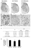
Melatonin ameliorates loss of motor neurons and atrophy of the spinal cord in mSOD1G93A ALS mice. Mice were sacrificed at the age of 120 days, and spinal cords were dissected after fixation. Serial sections of frozen tissue at the lumbar level were cut in the coronal plane at 50μm intervals and stained with Nissl substance. A–F, Representative Nissl-stained sections of spinal cord revealed the gross cross-sectional area (A–C) and motor-neuron content (D–F) for melatonin- and vehicle-treated ALS mice as well as for age-matched wild-type animals. Compared with wild-type littermate controls (A, D), gross spinal cord atrophy and ventral motor neuron loss were observed in vehicle-treated ALS mice (B, E), both of which were reduced by daily administration of 30 mg/kg melatonin (i.p.) (C, F). Scale bars in panels A–C and D–F correspond to 300 μm and 50 μm, respectively. G, Motor neuron counts and atrophy of gross areas grey matter, white matter and total area of spinal cord at the lumbar level were significantly rescued in melatonin-treated ALS mice. n = 5/group; ###, P < 0.001 vs. wild-type mice; **, P < 0.01; ***, P < 0.001 vs. vehicle-treated ALS mice. H, systemic injection of melatonin restored decreased levels of endogenous melatonin in the spinal cord of ALS mice. All values of intra-spinal melatonin levels were normalized to control values in wild-type mice. n = 6/group; #, P < 0.05 vs. wild-type mice; *, P < 0.05 vs. vehicle-treated ALS mice.
Figure 3
Figure 3
Melatonin inhibits release of apoptogenic factors from mitochondria and suppresses activation of caspases in mSOD1G93A ALS mice. 30 mg/kg of melatonin was administered by daily i.p. injection beginning at 6 weeks of age until the end of life. At the age of 120 days, mice were sacrificed and their spinal cords were removed. Cytosolic fractions or total lysates were prepared by homogenization and centrifugation as described in Materials and Methods. A, Samples of the cytosolic fractions were analyzed by western blotting using antibodies to cytochrome c and Smac (n = 4). B, Samples of total lysate were analyzed by western blotting with antibodies to pro- and active caspase-3, caspase-9 (n = 4). β-actin staining was used as an internal loading control. Each lane in these blots represents a different mouse. The bar graphs to the right are generated by densitometry. #, P < 0.05; ##, P < 0.01 vs. wild-type mice; *, P < 0.05 vs. vehicle-treated ALS mice.
Figure 4
Figure 4
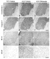
Melatonin decreases cytochrome c release, capase-3 activation, and gliosis in mSOD1G93A ALS mice. Mice were sacrificed at the age of 120 days, and their spinal cords were removed after fixation. 20-μm coronal cryosections were prepared and immunostained with antibodies to cytochrome c (A–C), activated caspase-3 (D–F), RCA-1 (G–I) or GFAP (J–L) (dark stained). Cytochrome c and activated caspase-3 are key mediators in mitochondria-related cell death pathways in ALS. The increased RCA-1 or GFAP expression corresponds to severity of microglia or astrocyte activation. The background immunostaining in the tissue samples are different as a consequence of cell arbors in the neuropil, where one would observe little of no background staining in wild-type mice. Immunostaining results demonstrated that cytochrome c release, capase-3 activation, and levels of reactive RCA-1 and GFAP increased in the vehicle-treated ALS mice (B, E, H, K) compared with wild-type mice (A, D, G, J). These pathophysiologic changes were reduced by treating the ALS mice with melatonin (C, F, I, L). Scale bars in (A–I) and (J–L) correspond to 150 μm and 50 μm, respectively.
Figure 5
Figure 5
Melatonin inhibits Rip2 overexpression and therefore prevents activation of caspase-1 in mSOD1G93A ALS mice. 30 mg/kg of melatonin was administered by daily i.p. injection beginning at 6 weeks of age until the end of life. At the age of 120 days, mice were sacrificed and their spinal cords were removed. Samples of total lysate fractions were analyzed by western blotting with antibodies to caspase-1 and Rip2 (wild-type groups, n = 4; ALS groups, n = 5). β-actin was used as an internal loading control. Each lane in the blots represents a different mouse. The bar graphs to the right are generated by densitometry. ##, P < 0.01 vs. wild-type mice; *, P < 0.05; **, P < 0.01 vs. vehicle-treated ALS mice.
Figure 6
Figure 6
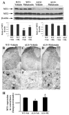
A significant down-regulated expression of MT1 in the spinal cord of ALS mSOD1G93A mice, and melatonin treatment protects against the depletion of MT1. A, Melatonin rescues MT1 expression in ALS mice. Samples of total lysates were analyzed by western blotting with antibodies to melatonin receptors MT1 and MT2 (wild-type groups, n = 4; ALS groups, n = 5). Each lane in the blots represents a different sample. The bar graphs are generated by densitometry. B–G, H, MT1 expression and distribution in the spinal cord were analyzed by immunostaining. 20-μm coronal cryosections were prepared and immunostained with antibodies to MT-1 in wild-type mice (B, E), vehicle (C, F), and melatonin (D, G) treated ALS mice. Scale bars in (B–D) and (E–G) correspond to 150 μm and 50 μm, respectively. n = 5/group; #, P < 0.05; ##, P < 0.01 vs. wild-type mice. *, P < 0.05; **, P < 0.01 vs. vehicle-treated ALS mice.

References

    1. Bensimon G, et al. A controlled trial of riluzole in amyotrophic lateral sclerosis. ALS/Riluzole Study Group. N Engl J Med. 1994;330:585–91. - PubMed
    1. Caballero B, et al. Favorable effects of a prolonged treatment with melatonin on the level of oxidative damage and neurodegeneration in senescence-accelerated mice. J Pineal Res. 2008;45:302–11. - PubMed
    1. Cleveland DW, Rothstein JD. From Charcot to Lou Gehrig: deciphering selective motor neuron death in ALS. Nat Rev Neurosci. 2001;2:806–19. - PubMed
    1. Das A, et al. Cytoprotective effects of melatonin on C6 astroglial cells exposed to glutamate excitotoxicity and oxidative stress. J Pineal Res. 2008;45:117–24. - PMC - PubMed
    1. Drachman DB, et al. Cyclooxygenase 2 inhibition protects motor neurons and prolongs survival in a transgenic mouse model of ALS. Ann Neurol. 2002;52:771–8. - PubMed

Publication types