

Deformed grids for single-particle cryo-electron microscopy of specimens exhibiting a preferred orientation
- PMID: 23537848
- PMCID: PMC3665629
- DOI: 10.1016/j.jsb.2013.03.005
Deformed grids for single-particle cryo-electron microscopy of specimens exhibiting a preferred orientation
Abstract
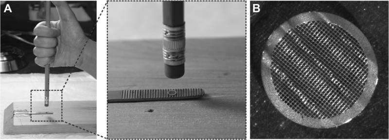
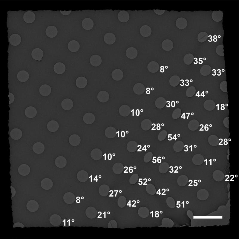
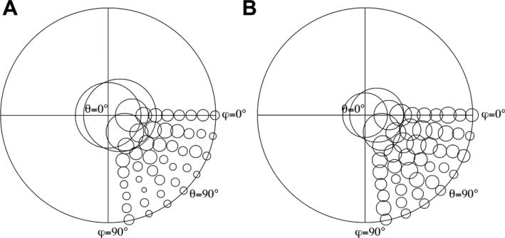
For biological samples showing a preferred orientation on the carbon support film of an electron microscope (EM) grid, accurate three-dimensional (3D) reconstructions by single-particle cryo-EM require data collection in which the specimen grids are tilted in the microscope, to obtain adequate numbers of particles that cover the high-degree angular distribution. However, image drift caused by the electron beam interacting with the cryo specimen becomes severe when grids are tilted to high angles (>30°). We produced deformed grids by applying a deliberate mechanical deformation to EM grids containing a thin carbon film supported by a thick holey carbon film. We applied cryo-EM using deformed grids to the isolated cardiac ryanodine receptor, an ion channel complex known to assume a preferred orientation on the carbon support film. These grids contained more particles having high Euler angle orientations without the need to tilt the specimen grids. Meanwhile, the drifting that was apparent in the images was reduced from that typical of images from tilted regular EM grids. This was achieved by imaging particles in holes close to the deformed areas, where carbon films were locally bent, offering planes of inclination with various angles. The deformed grids improve the efficiency and quality of data collection for single-particle cryo-EM of samples showing a limited range of orientations.
Copyright © 2013 Elsevier Inc. All rights reserved.
Figures
References
-
- Deatherage JF, Taylor KA, Amos LA. Three-dimensional arrangement of the cell wall protein of Sulfolobus acidocaldarius. J. Mol. Biol. 1983;167:823–848. - PubMed
-
- Downing KH, McCartney MR, Glaeser RM. Experimental characterization and mitigation of specimen charging on thin films with one conducting layer. Microsc. Microanal. 2004;10:783–789. - PubMed
-
- Frank J, Radermacher M, Penczek P, Zhu J, Li Y, Ladjadj M, Leith A. SPIDER and WEB: Processing and visualization of images in 3D electron microscopy and related fields. J. Struct. Biol. 1996;116:190–199. - PubMed
-
- Fujiyoshi Y, Mizusaki T, Morikawa K, Yamagishi H, Aoki Y, Kihara H, Harada Y. Development of a superfluid helium stage for high-resolution electron microscopy. Ultramicroscopy. 1991;38:241–251.
Publication types
MeSH terms
Substances
Grants and funding
LinkOut - more resources
Full Text Sources
Other Literature Sources
