

The association of fetal cerebrovascular resistance with early neurodevelopment in single ventricle congenital heart disease
- PMID: 23537971
- PMCID: PMC3644301
- DOI: 10.1016/j.ahj.2012.11.013
The association of fetal cerebrovascular resistance with early neurodevelopment in single ventricle congenital heart disease
Abstract
Background: Children with congenital heart disease are at risk for impaired neurodevelopment (ND). We investigated the association of fetal cerebrovascular resistance with ND in patients with single ventricle lesions.
Methods: In the Single Ventricle Reconstruction (SVR) and Infant Single Ventricle trials, 14-month ND was assessed using the Bayley Scales of Infant Development II. We investigated associations between ND scores and fetal middle cerebral artery pulsatility index (MCA-PI) z-scores, a Doppler-derived estimate of cerebrovascular resistance in a subset of those infants.
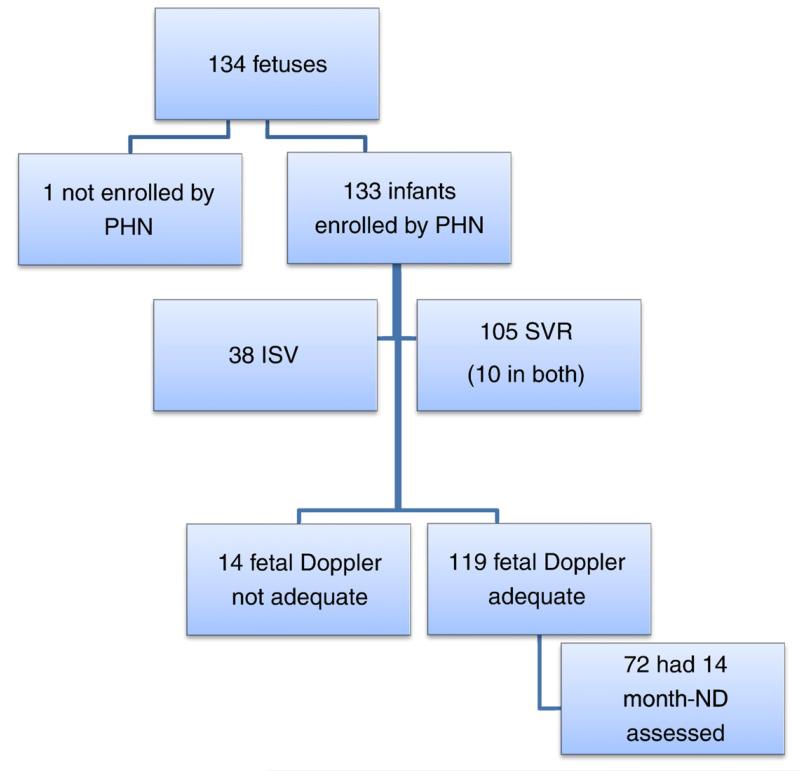
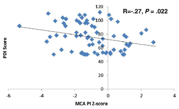
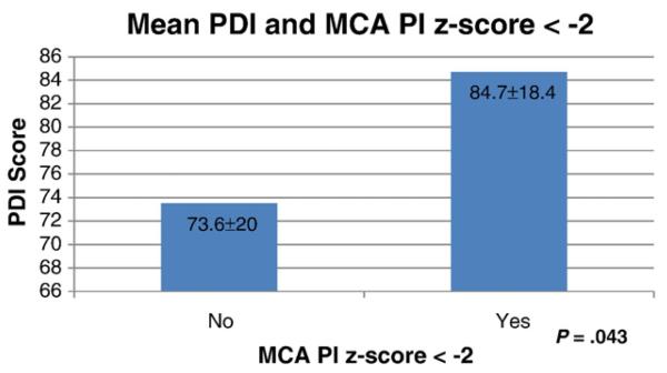
Results: Neurodevelopment assessments were performed at age 14.3 ± 1 months in 170 (74%) of 230 Infant Single Ventricle and 321 (58%) of 555 SVR subjects. Fetal echocardiographic data were available in 119 subjects, 72 (61%) of which had ND testing. Mean Psychomotor Development Index (PDI) (76 ± 20) and Mental Development Index (MDI) (89 ± 17) scores were lower than normative means (100 ± 15, P < .001). Mean MCA-PI z-score was -0.95 ± 1.52. Middle cerebral artery pulsatility index z-score correlated negatively with PDI (r = -0.27, P = .02) but was not associated with MDI. When MCA-PI z-score was added to a multivariable model controlling for factors identified in the SVR trial to predict PDI, the percentage of explained variation increased from 23% to 30%, and MCA-PI z-score remained an independent predictor (r = -3.864, P = .03). Middle cerebral artery pulsatility index z-score was not an independent predictor in a model adjusting for site.
Conclusions: Among fetuses with single ventricle anomalies, lower cerebrovascular resistance was associated with higher ND scores. This relationship is opposite to that observed with advanced intrauterine growth retardation and may represent a unique ability of these congenital heart disease fetuses to compensate for diminished cerebral oxygen delivery.
Trial registration: ClinicalTrials.gov NCT00115934.
Copyright © 2013 Mosby, Inc. All rights reserved.
Figures
References
-
- Tabbutt S, Nord AS, Jarvik GP, et al. Neurodevelopmental outcomes after staged palliation for hypoplastic left heart syndrome. Pediatr. 2008;121(3):476–83. - PubMed
-
- Goldberg CS, Schwartz EM, Brunberg JA, et al. Neurodevelopmental outcome of patients after the fontan operation: a comparison between children with hypoplastic left heart syndrome and other functional single ventricle lesions. J Pediatr. 2000;137(5):646–52. - PubMed
-
- Wernovsky G, Shillingford AJ, Gaynor JW. Central nervous system outcomes in children with complex congenital heart disease. Curr Op Cardiol. 2005;20(2):94–9. - PubMed
-
- Peeters LL, Sheldon RE, Jones MD, Jr, et al. Blood flow to fetal organs as a function of arterial oxygen content. Am J Obstet Gynecol. 1979;135(5):637–46. - PubMed
-
- Dubiel M, Gunnarsson GO, Gudmundsson S. Blood redistribution in the fetal brain during chronic hypoxia. Ultrasound Obstet Gynecol. 2002;20(2):117–21. - PubMed
Publication types
MeSH terms
Substances
Associated data
Grants and funding
LinkOut - more resources
Full Text Sources
Other Literature Sources
Medical
Research Materials
Miscellaneous
