

Comparison of 10 TTP and Tmax estimation techniques for MR perfusion-diffusion mismatch quantification in acute stroke
- PMID: 23538410
- PMCID: PMC7965638
- DOI: 10.3174/ajnr.A3460
Comparison of 10 TTP and Tmax estimation techniques for MR perfusion-diffusion mismatch quantification in acute stroke
Abstract
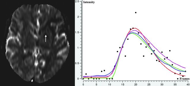
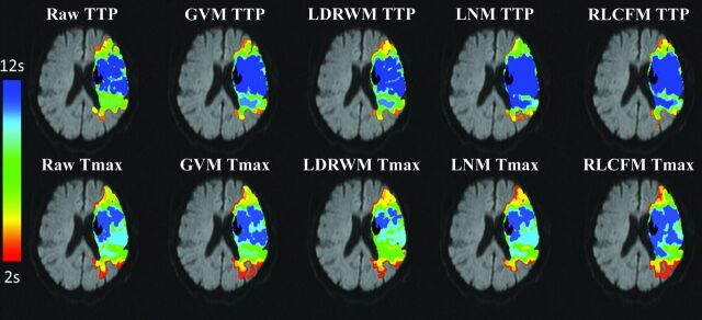
Background and purpose: The mismatch between lesions identified in perfusion- and diffusion-weighted MR imaging is typically used to identify tissue at risk of infarction in acute stroke. The purpose of this study was to analyze the variability of mismatch volumes resulting from different time-to-peak or time-to-maximum estimation techniques used for hypoperfused tissue definition.
Materials and methods: Data of 50 patients with middle cerebral artery stroke and intracranial vessel occlusion imaged within 6 hours of symptom onset were analyzed. Therefore, 10 different TTP/Tmax techniques and delay thresholds between +2 and +12 seconds were used for calculation of perfusion lesions. Diffusion lesions were semiautomatically segmented and used for mismatch quantification after registration.
Results: Mean volumetric differences up to 40 and 100 mL in individual patients were found between the mismatch volumes calculated by the 10 TTP/Tmax estimation techniques for typically used delay thresholds. The application of typical criteria for the identification of patients with a clinically relevant mismatch volume resulted in different mismatch classifications in ≤24% of all cases, depending on the TTP/Tmax estimation method used.
Conclusions: High variations of tissue-at-risk volumes have to be expected when using different TTP/Tmax estimation techniques. An adaption of different techniques by using correction formulas may enable more comparable study results until a standard has been established by agreement.
Figures
Similar articles
-
Comparison of computed tomography perfusion and magnetic resonance imaging perfusion-diffusion mismatch in ischemic stroke.Stroke. 2012 Oct;43(10):2648-53. doi: 10.1161/STROKEAHA.112.660548. Epub 2012 Aug 2. Stroke. 2012. PMID: 22858726
-
Refining the perfusion-diffusion mismatch hypothesis.Stroke. 2005 Jun;36(6):1153-9. doi: 10.1161/01.str.0000166181.86928.8b. Stroke. 2005. PMID: 15914768 Clinical Trial.
-
Visual assessment of magnetic resonance imaging perfusion lesions in a large patient group.Clin Neuroradiol. 2012 Dec;22(4):305-13. doi: 10.1007/s00062-012-0143-4. Epub 2012 Apr 8. Clin Neuroradiol. 2012. PMID: 22484907
-
Mismatch of delayed perfusion volume between TTP and Tmax map of perfusion MRI.Clin Imaging. 2016 Jan-Feb;40(1):63-7. doi: 10.1016/j.clinimag.2015.10.005. Epub 2015 Oct 21. Clin Imaging. 2016. PMID: 26597103
-
Automated estimation of salvageable tissue: Comparison with expert readers.J Magn Reson Imaging. 2016 Jan;43(1):220-8. doi: 10.1002/jmri.24963. Epub 2015 Jun 2. J Magn Reson Imaging. 2016. PMID: 26036930
Cited by
-
Core and penumbra estimation using deep learning-based AIF in association with clinical measures in computed tomography perfusion (CTP).Insights Imaging. 2023 Sep 29;14(1):161. doi: 10.1186/s13244-023-01472-z. Insights Imaging. 2023. PMID: 37775600 Free PMC article.
-
Prediction of tissue outcome in acute ischemic stroke based on single-phase CT angiography at admission.Front Neurol. 2024 Mar 19;15:1330497. doi: 10.3389/fneur.2024.1330497. eCollection 2024. Front Neurol. 2024. PMID: 38566856 Free PMC article.
-
Impact of Severe Extracranial ICA Stenosis on MRI Perfusion and Diffusion Parameters in Acute Ischemic Stroke.Front Neurol. 2014 Dec 5;5:254. doi: 10.3389/fneur.2014.00254. eCollection 2014. Front Neurol. 2014. PMID: 25538674 Free PMC article.
-
Mapping causal functional contributions derived from the clinical assessment of brain damage after stroke.Neuroimage Clin. 2015 Aug 1;9:83-94. doi: 10.1016/j.nicl.2015.07.009. eCollection 2015. Neuroimage Clin. 2015. PMID: 26448908 Free PMC article.
-
Stroke Lesion Segmentation in FLAIR MRI Datasets Using Customized Markov Random Fields.Front Neurol. 2019 May 24;10:541. doi: 10.3389/fneur.2019.00541. eCollection 2019. Front Neurol. 2019. PMID: 31178820 Free PMC article.
References
-
- Muir KW, Buchan A, von Kummer R, et al. . Imaging of acute stroke. Lancet Neurol 2006;5:755–68 - PubMed
-
- Butcher KS, Parsons M, MacGregor L, et al. . Refining the perfusion-diffusion mismatch hypothesis. Stroke 2005;36:1153–59 - PubMed
-
- Neumann-Haefelin T, Wittsack HJ, Wenserski F, et al. . Diffusion- and perfusion-weighted MRI. The DWI/PWI mismatch region in acute stroke. Stroke 1999;30:1591–97 - PubMed
-
- Sobesky J, Zaro Weber O, Lehnhardt FG, et al. . Which time-to-peak threshold best identifies penumbral flow? A comparison of perfusion-weighted magnetic resonance imaging and positron emission tomography in acute ischemic stroke. Stroke 2004;35:2843–47 - PubMed
-
- Thomalla G, Schwark C, Sobesky J, et al. . Outcome and symptomatic bleeding complications of intravenous thrombolysis within 6 hours in MRI-selected stroke patients: comparison of a German multicenter study with the pooled data of ATLANTIS, ECASS, and NINDS tPA trials. Stroke 2006;37:852–58 - PubMed
Publication types
MeSH terms
LinkOut - more resources
Full Text Sources
Other Literature Sources
Medical