

Diffuse hemolymphangioma of the rectum: a report of a rare case
- PMID: 23538679
- PMCID: PMC3602512
- DOI: 10.3748/wjg.v19.i9.1494
Diffuse hemolymphangioma of the rectum: a report of a rare case
Abstract
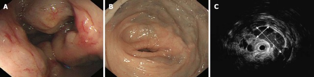
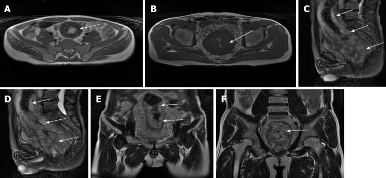
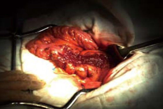
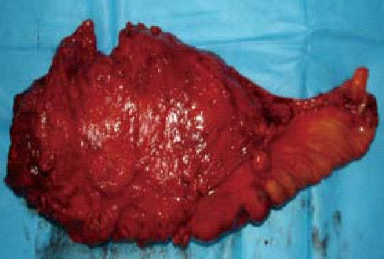
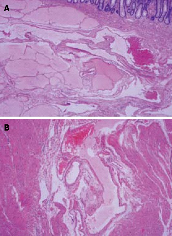
Intestinal hemolymphangioma is a rare vascular and lymphatic malformation and is manifested as anaemia and recurrent alimentary tract hemorrhage. Few cases of hemolymphangioma occurring in small intestine, spleen, esophagus and other organs have been reported. We herein report a case of a 37-year-old man with severe rectal bleeding. Digital examination revealed nodular mucosa. No rectal mass was palpated, but bleeding in the ampulla was detected. Colonoscopy revealed an extensive hypervascular submucosal lesion arising from the rectosigmoid junction colon to the distal edge of the anus. Endoscopic ultrasonography demonstrated an extensive anechoic mass with clear edge. Magnetic resonance imaging (MRI) showed a significant thickness of the rectal wall, extending to the distal edge of the anus, with a narrowing lumen. A sphincter-saving rectal surgery was performed. Due to a lack of knowledge of the clinical, endoscopic and radiological features, preoperative recognition of hemolymphangioma is not easy. Computed tomography and MRI are helpful in confirming the diagnosis, and defining the extent and invasion of the lesion. For the low malignant potential tumors, a sphincter-saving rectal surgery is recommended after a full evaluation of the tumor.
Keywords: Hemolymphangioma; Rectal bleeding; Rectum.
Figures
References
-
- Bethouart M, Houcke M, Proye C, Linquette M. Hepatosplenic hemolymphangioma. Lille Med. 1980;25:288–290. - PubMed
-
- Canavese F, Cortese MG, Proietti L, Costantino S, Rosina M, Nangeroni M, Defilippi C, Di Rosa GP. Bulky-pedunculated hemolymphangioma of the esophagus: rare case in a two-years old girl. Eur J Pediatr Surg. 1996;6:170–172. - PubMed
-
- Nataf P, Mestiri T, Martin de Lasalle E, Benomar M, Gandjbakhch I, Cabrol C. Pericardial hemolymphangioma. Apropos of a case. Arch Mal Coeur Vaiss. 1988;81:1137–1140. - PubMed
Publication types
MeSH terms
LinkOut - more resources
Full Text Sources
Other Literature Sources
