. 2013 Nov;27(11):2177-86.
doi: 10.1038/leu.2013.91. Epub 2013 Mar 29.

Global H3K4me3 genome mapping reveals alterations of innate immunity signaling and overexpression of JMJD3 in human myelodysplastic syndrome CD34+ cells

Affiliations

Global H3K4me3 genome mapping reveals alterations of innate immunity signaling and overexpression of JMJD3 in human myelodysplastic syndrome CD34+ cells

Y Wei et al. Leukemia. 2013 Nov.

Abstract

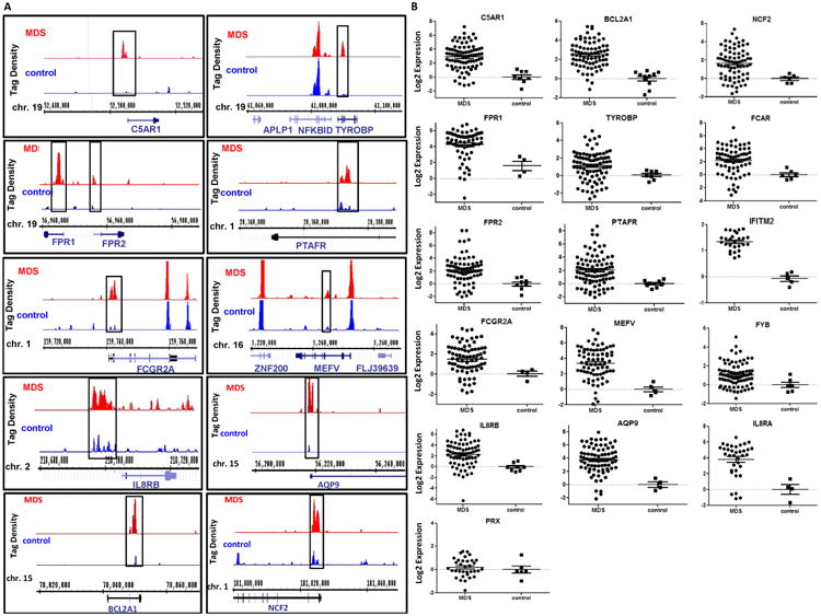
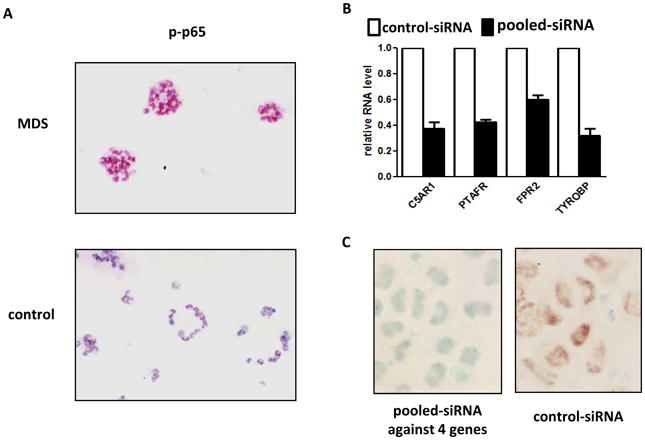
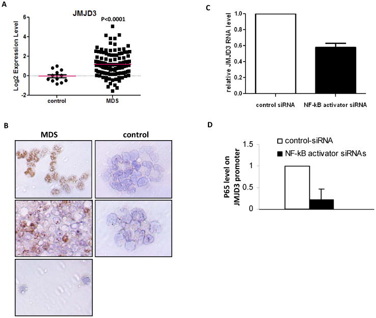
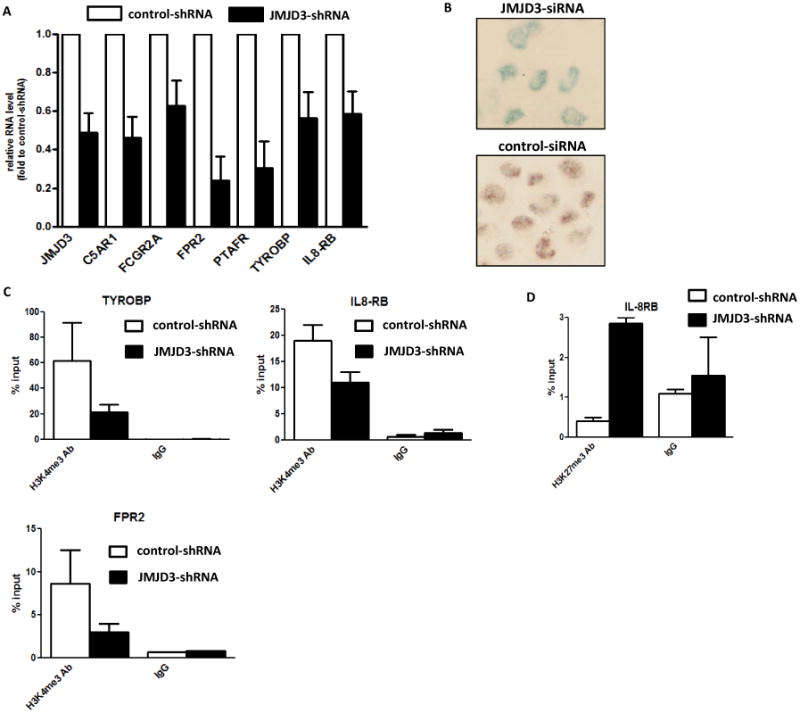
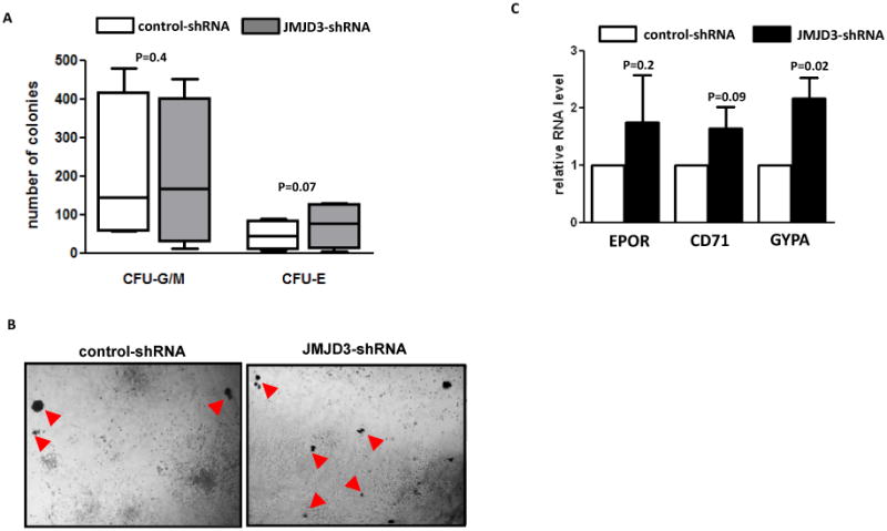
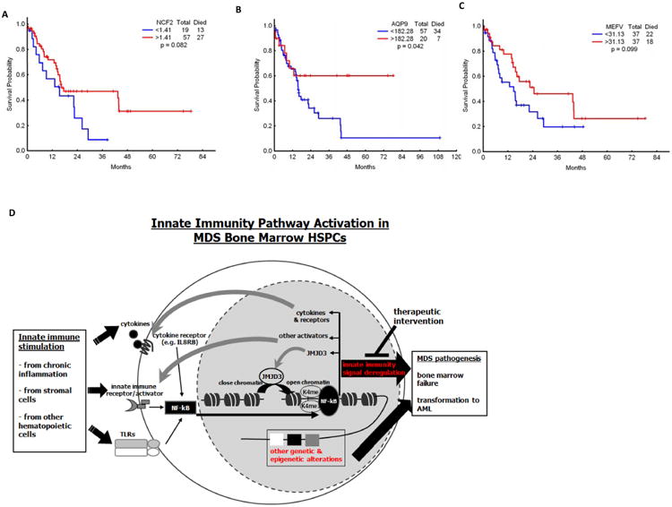
The molecular bases of myelodysplastic syndromes (MDS) are not fully understood. Trimethylated histone 3 lysine 4 (H3K4me3) is present in promoters of actively transcribed genes and has been shown to be involved in hematopoietic differentiation. We performed a genome-wide H3K4me3 CHIP-Seq (chromatin immunoprecipitation coupled with whole genome sequencing) analysis of primary MDS bone marrow (BM) CD34+ cells. This resulted in the identification of 36 genes marked by distinct higher levels of promoter H3K4me3 in MDS. A majority of these genes are involved in nuclear factor (NF)-κB activation and innate immunity signaling. We then analyzed expression of histone demethylases and observed significant overexpression of the JmjC-domain histone demethylase JMJD3 (KDM6b) in MDS CD34+ cells. Furthermore, we demonstrate that JMJD3 has a positive effect on transcription of multiple CHIP-Seq identified genes involved in NF-κB activation. Inhibition of JMJD3 using shRNA in primary BM MDS CD34+ cells resulted in an increased number of erythroid colonies in samples isolated from patients with lower-risk MDS. Taken together, these data indicate the deregulation of H3K4me3 and associated abnormal activation of innate immunity signals have a role in the pathogenesis of MDS and that targeting these signals may have potential therapeutic value in MDS.


Conflict of interest statement

Conflict of Interest: None of the authors have any conflict of interest with the data presented here.

Figures

Figure 1
Figure 1. H3K4me3 CHIP-Seq in primary MDS bone marrow CD34+ cells
(a) Representative examples of H3K4me3 identified by CHIP-Seq in 11 genes in MDS (top) and control (bottom) BM CD34+ cells. Comparison between MDS and control samples indicate increased levels of H3K4me3 in the promoters of these genes in MDS CD34+ cells. (b) Logarithmic representation of Q-RTPCR results of 16 genes identified by CHIP-Seq in MDS and control CD34+ cells. These results confirm increased RNA expression in genes marked by higher levels of promoter H3K4me3 in MDS.
Figure 2
Figure 2. Genes identified by CHIP-Seq are involved in NF-kB activation in MDS CD34+ cells
(a) Immuno-histochemical analysis of phospho-p65 in MDS (Top panel) and controls (Bottom panel) bone marrow CD34+ cells. (b) Knock-down of C5AR1, FPR2, TYROBP and PTAFR in OCI-AML3 cells. (c) Immuno-histochemical analysis of phospho-p65 in the OCI-AML3 cells after knock-down of the 4 genes described above (Left panel) in comparison to control siRNA transfected cells (Right panel).
Figure 3
Figure 3. JMJD3 expression in MDS BM CD34+ cells
(a) Logarithmic representation of JMJD3 Q-RTPCR results in MDS and control CD34+ cells in 121 MDS CD34+ samples. (b) Immuno-histochemical analysis of JMJD3 in cytospins of MDS (Left 3 panels: Top 2 with strong JMJD3 staining and Bottom 1 with weak JMJD3 staining) and controls (Right 2 panels: both with weak JMJD3 staining) bone marrow CD34+ cells. (c) Q-RTPCR analysis of JMJD3 RNA expression in OCI-AML3 cells transfected with siRNAs targeting C5AR1, FPR2, TYROBP and PTAFR or controls. (d) p65 CHIP-PCR analysis of JMJD3 promoter in the OCI-AML3 cells transfected with the siRNAs targeting C5AR1, FPR2, TYROBP and PTAFR or controls.
Figure 4
Figure 4. JMJD3 is involved in transcriptional regulation of genes identified by CHIP-Seq
(a) Q-RTPCR analysis of RNA expression of 6 genes involved in NF-kB activation in OCI-AML3 cells after JMJD3 knock-down. (b) Immuno-histochemical analysis of phospho-p65 in the OCI-AML3 cells with JMJD3 knock-down (Top panel) and in control cells (Bottom panel). (c) H3K4me3 CHIP-PCR analysis of IL8RB, TYROBP, and FPR2 in the OCI-AML3 cells after JMJD3 knock-down. (d) H3K27me3 CHIP-PCR analysis of IL8RB in the OCI-AML3 cells after JMJD3 knock-down.
Figure 5
Figure 5. Effect of JMJD3 shRNA transduction in cultured MDS bone marrow CD34+ cells
(a) Numbers of myeloid colonies (CFU-G/M) and erythroid colonies (CFU-E) formed in methocult culture two weeks after transduction of JMJD3-shRNA and control shRNA in BM CD34+ cells isolated from patients with lower-risk MDS (low-risk and intermediate-1). (b) Representative microphotographs of colonies formed in methocult plates after transduction of control shRNA (Left panel) and JMJD3-shRNA (Right panel). Red arrows point to CFU-E. (c) Q-RTPCR analysis of the RNA levels of CD71, EPOR and GYPA in cells collected from total colonies after shRNA transduction and methocult assays.
Figure 6
Figure 6. Survival impact of gene expression in MDS and proposed model of implications of innate immunity signaling in MDS
(a-c) Effect of mRNA expression of NCF2, AQP9 and MEFV on survival of patients with MDS. (d) Innate immunity stimulation derived from stroma, chronic inflammation or hematopoietic cells either in a para or autocrine fashion result in NF-kB activation in MDS CD34+ cells. NF-kB activation then triggers expression of multiple other effectors such as cytokines and importantly JMJD3. JMJD3 is a histone demethylase that contributes to the perpetuation of innate immunity signal and NF-kB activation. Signaling via this pathway probably cooperates with other known genetic and epigenetic lesions known to occur in MDS cells and contribute to bone marrow failure and transformation to AML that characterized MDS. Further analysis of this pathway could result in the development of inhibitors with therapeutic potential.

References

    1. Tefferi A, Vardiman JW. Myelodysplastic syndromes. N Engl J Med. 2009;361(19):1872–85. - PubMed
    1. Garcia-Manero G, Fenaux P. Hypomethylating Agents and Other Novel Strategies in Myelodysplastic Syndromes. J Clin Oncol. 2011;29(10):516–23. - PMC - PubMed
    1. Cutler CS, Lee SJ, Greenberg P, Deeg HJ, Perez WS, Anasetti C, et al. A decision analysis of allogeneic bone marrow transplantation for the myelodysplastic syndromes: delayed transplantation for low-risk myelodysplasia is associated with improved outcome. Blood. 2004;104(2):579–85. - PubMed
    1. Slape C, Lin YW, Hartung H, Zhang Z, Wolff L, Aplan PD. NUP98-HOX translocations lead to myelodysplastic syndrome in mice and men. J Natl Cancer Inst Monogr. 2008;(39):64–8. Epub 2008/07/24. - PMC - PubMed
    1. Schanz J, Steidl C, Fonatsch C, Pfeilstocker M, Nosslinger T, Tuechler H, et al. Coalesced multicentric analysis of 2,351 patients with myelodysplastic syndromes indicates an underestimation of poor-risk cytogenetics of myelodysplastic syndromes in the international prognostic scoring system. J Clin Oncol. 2011;29(15):1963–70. - PMC - PubMed

MeSH terms