. 2013 Jul;11(7):714-23.
doi: 10.1158/1541-7786.MCR-12-0668. Epub 2013 Mar 28.

Oncogenic MUC1-C promotes tamoxifen resistance in human breast cancer

Affiliations

Oncogenic MUC1-C promotes tamoxifen resistance in human breast cancer

Akriti Kharbanda et al. Mol Cancer Res. 2013 Jul.

Abstract

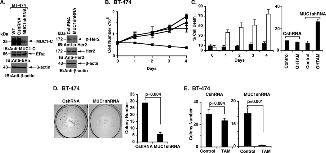
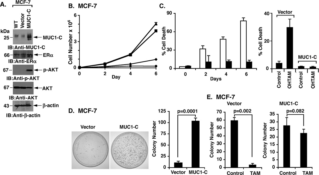
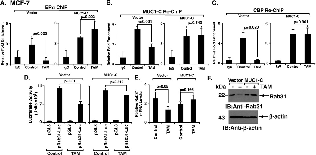
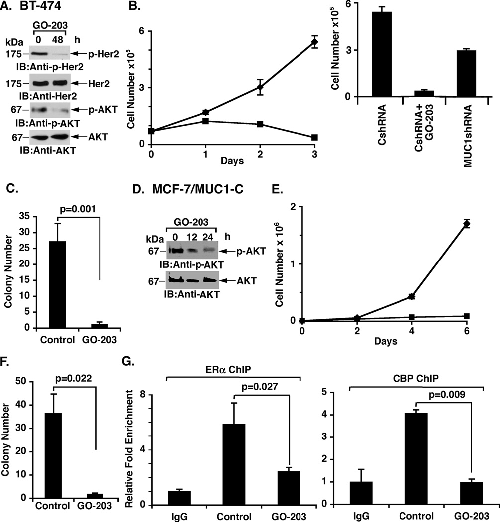
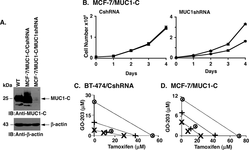
Tamoxifen resistance of estrogen receptor-positive (ER+) breast cancer cells has been linked in part to activation of receptor tyrosine kinases, such as HER2, and the PI3K-AKT pathway. Mucin 1 (MUC1) is aberrantly overexpressed in about 90% of human breast cancers, and the oncogenic MUC1-C subunit is associated with ERα. The present studies using HER2 overexpressing BT-474 breast cancer cells, which are constitutively resistant to tamoxifen, demonstrate that silencing MUC1-C is associated with (i) downregulation of p-HER2 and (ii) sensitivity to tamoxifen-induced growth inhibition and loss of clonogenic survival. In contrast, overexpression of MUC1-C in tamoxifen-sensitive MCF-7 breast cancer cells resulted in upregulation of p-AKT and tamoxifen resistance. We show that MUC1-C forms complexes with ERα on the estrogen-responsive promoter of Rab31 and that MUC1-C blocks tamoxifen-induced decreases in ERα occupancy. MUC1-C also attenuated tamoxifen-induced decreases in (i) recruitment of the coactivator CREB binding protein, (ii) Rab31 promoter activation, and (iii) Rab31 mRNA and protein levels. The importance of MUC1-C is further supported by the demonstration that targeting MUC1-C with the cell-penetrating peptide inhibitor, GO-203, sensitized tamoxifen-resistant cells to tamoxifen treatment. Moreover, we show that targeting MUC1-C in combination with tamoxifen is highly synergistic in the treatment of tamoxifen-resistant breast cancer cells. Combined, these findings indicate that MUC1-C contributes to tamoxifen resistance.


Conflict of interest statement

Conflict of Interest: D. Kufe is a founder of Genus Oncology and holds equity in the company. The other authors disclosed no potential conflicts of interest.

Figures

Figure 1
Figure 1. Resistance of HER2-overexpressing BT-474 cells to tamoxifen is conferred by MUC1-C expression
A. Lysates from wild-type (WT) BT-474 cells, BT-474/CshRNA and BT-474/MUC1shRNA cells were immunoblotted with the indicated antibodies (left and right). B. Control BT-474/CshRNA (squares) and BT-474/MUC1shRNA (diamonds) cells were left untreated. BT-474/CshRNA (triangles) and BT-474/MUC1shRNA (circles) were also treated with 5 µM tamoxifen on days 0 and 2. Cell number is expressed as the mean±SD of three replicates. C. BT-474/CshRNA (solid bars) and BT-474/MUC1shRNA (open bars) cells were treated with 5 µM tamoxifen (left) or 10 nM OHTAM (right) on days 0 and 2. The results (mean±SD of three replicates) are expressed as the percentage of cell death as determined by trypan blue staining on the indicated days (left) or on day 4 (right). D. BT-474/CshRNA and BT-474/MUC1shRNA cells were seeded at 1000 cells/well (6-well plate), grown for 14 days and then stained with crystal violet (left). Colony number (>30 cells) is expressed as the mean±SD of three replicates (right). E. BT-474/CshRNA (1000 cells/well; left) and BT-474/MUC1shRNA (2000 cells/well; right) cells were seeded in 6-well plates and left untreated (Control) or treated with 5 µM tamoxifen (TAM) every other day for 14 days. Colony number (>30 cells) is expressed as the mean±SD of three replicates.
Figure 2
Figure 2. Overexpression of MUC1-C in MCF-7 cells is associated with tamoxifen resistance
A. Lysates from wild-type (WT) MCF-7 cells, MCF-7/vector and MCF-7/MUC1-C cells were immunoblotted with the indicated antibodies. B. Control MCF-7/vector (diamonds) and MCF-7/MUC1-C (triangles) cells were left untreated. MCF-7/vector (circles) and MCF-7/MUC1-C (squares) cells were also treated with 5 µM tamoxifen on days 0, 2 and 4. Cell number is expressed as the mean±SD of three replicates. C. MCF-7/vector (open bars) and MCF-7/MUC1-C (solid bars) cells were treated with 5 µM tamoxifen on days 0, 2 and 4 (left) or 10 nM OHTAM on days 0 and 2 (right). The results (mean±SD of three replicates) are expressed as the percentage of cell death as determined by trypan blue staining on the indicated days (left) or on day 4 (right). D. MCF-7/vector and MCF-7/MUC1-C cells were seeded at 500 cells/well (6-well plate), grown for 7 days and then stained with crystal violet (left). Colony number (>30 cells) is expressed as the mean±SD of three replicates (right). E. MCF-7/vector (2000 cells/well; left) and MCF-7/MUC1-C (500 cells/well; right) cells were seeded in 6-well plates and left untreated (Control) or treated with 5 µM tamoxifen (TAM) every other day for 7 days. Colony number (>30 cells) is expressed as the mean±SD of three replicates.
Figure 3
Figure 3. MCF-7 cells overexpressing MUC1-C are estrogen-independent
A. MCF-7/vector (squares) and MCF-7/MUC1-C (circles) were seeded at 1 × 10⁴ cells/ml in IMEM/CSS medium for the indicated number of days. The results are expressed as the cell number × 106/ml (mean±SD of three replicates). B. MCF-7/vector (open bars) and MCF-7/MUC1-C (solid bars) cells were seeded in IMEM/CSS medium for the indicated number of days. The results are expressed as the percentage of cell death (mean±SD of three replicates) as determined by trypan blue staining. C. MCF-7/vector and MCF-7/MUC1-C cells were seeded at 500 cells/well (6-well plate), grown for 7 days in IMEM/CSS medium and then stained with crystal violet (left). Colony number (>30 cells) is expressed as the mean±SD of three replicates (right).
Figure 4
Figure 4. MUC1-C blocks the effects of tamoxifen on occupancy and activation of the Rab31 promoter
A–C. MCF-7/vector (left) and MCF-7/MUC1-C (right) cells were left untreated (Control) or treated with 5 µM tamoxifen for 2 days. A. Soluble chromatin was precipitated with anti-ERα or a control IgG. The final DNA samples were analyzed for Rab31 promoter estrogen-responsive element (ERE) or control region (CR) sequences (16). The results (mean+SD of three determinations) are expressed as the relative fold enrichment compared to that obtained with the IgG control. B and C. In re-ChIP experiments, the anti-ERα precipitates were released, reimmunoprecipitated with anti-MUC1-C (B) or anti-CBP (C) and a control IgG, and then analyzed for Rab31 promoter sequences. The results (mean±SD of three determinations) are expressed as the relative fold enrichment compared to that obtained with the IgG control. D. MCF-7/vector and MCF-7/MUC1-C cells were transfected with the pGL3 vector or pRab31-Luc and Renilla plasmid as an internal control. The cells were then left untreated (Control) or treated with 5 µM tamoxifen for 2 days and then assayed for luciferase activity. The results are expressed as the mean±SD of two experiments each performed with three determinations. E. MCF-7/vector and MCF-7/MUC1-C cells were left untreated (Control) or treated with 5 µM tamoxifen for 2 days. Rab31 and GAPDH mRNA levels were determined by qRT-PCR. The results (mean±SD of three determinations) are expressed as relative Rab31 mRNA levels as compared to that obtained for GAPDH. F. MCF-7/vector and MCF-7/MUC1-C cells were left untreated (Control) or treated with 5 µM tamoxifen for 2 days. Lysates from the indicated cells were immunoblotted with anti-Rab31 and anti-β-actin.
Figure 5
Figure 5. Tamoxifen-resistant cells are sensitive to MUC1-C inhibition
A. BT-474 cells were left untreated (Control) or treated with 5 µM GO-203 each day for 2 days. Lysates were immunoblotted with the indicated antibodies. B. BT-474/CshRNA cells were left untreated (diamonds) or treated with 5 µM GO-203 (squares) each day for the indicated days (left). The results (mean±SD of three replicates) are expressed as the viable cell number. BT-474/CshRNA and BT-474/MUC1shRNA cells were seeded at 5 × 10⁴ cells/well and left untreated (right). The BT-474/CshRNA cells were also treated with 5 µM GO-203 each day for 3 days. The results (mean±SD of three replicates) are expressed as the viable cell number on day 4. C. BT-474/CshRNA cells were seeded at 1000 cells/well in 6-well plates and left untreated (Control) or treated with 5 µM GO-203 each day for 7 days. Colony number (>30 cells) is expressed as the mean±SD of three replicates. D. MCF-7/MUC1-C cells were left untreated (Control) or treated with 5 µM GO-203 each day for 2 days. Lysates were immunoblotted with the indicated antibodies. E. MCF-7/MUC1-C cells were left untreated (diamonds) or treated with 5 µM GO-203 (squares) each day for the indicated days. The results (mean±SD of three replicates) are expressed as the viable cell number. F. MCF-7/MUC1-C cells were seeded at 200 cells/well in 6-well plates and left untreated (Control) or treated with 5 µM GO-203 each day for 7 days. Colony number (>30 cells) is expressed as the mean±SD of three replicates. G. MCF-7/MUC1-C cells were left untreated (Control) or treated with 5 µM GO-203 each day for 2 days. Soluble chromatin was precipitated with anti-ERα (left), anti-CBP (right) or a control IgG. The precipitates were analyzed for Rab31 promoter sequences. The results (mean±SD of three determinations) are expressed as the relative fold enrichment compared to that obtained with the IgG control.
Figure 6
Figure 6. Synergistic interaction between GO-203 and tamoxifen
A. MCF-7/MUC1-C cells were infected with lentiviruses expressing the control shRNA (CshRNA) or the MUC1shRNA. Lysates from wild-type (WT) MCF-7 cells, MCF-7/MUC1-C/CshRNA and MCF-7/MUC1-C/MUC1shRNA cells were immunoblotted with the indicated antibodies. B. MCF-7/MUC1-C/CshRNA (left) and MCF-7/MUC1-C/MUC1shRNA (right) cells were left untreated (triangles) or treated (circles) with 5 µM tamoxifen days 0 and 2. The results (mean±SD of three replicates) are expressed as viable cell number. C and D. BT-474/CshRNA (C) and MCF-7/MUC1-C (D) cells were treated with fixed IC50 ratios of (i) GO-203 alone on days 0, 1, 2, 3 and 4, (ii) tamoxifen alone on days 0, 2 and 4 and (iii) the GO-203/tamoxifen combination. For tamoxifen-resistant BT-474/CshRNA and MCF-7/MUC1-C cells, tamoxifen was used at the half-maximal inhibitory concentrations obtained for the tamoxifen-sensitive BT-474/MUC1shRNA and MCF-7/vector cells, respectively. The multiple effect-level isobologram analyses are shown for the ED50 (×), ED75 (+) and ED90 (◉) values.

References

    1. Osborne CK, Schiff R. Mechanisms of endocrine resistance in breast cancer. Annu Rev Med. 2011;62:233–247. - PMC - PubMed
    1. Musgrove EA, Sutherland RL. Biological determinants of endocrine resistance in breast cancer. Nat Rev Cancer. 2009;9(9):631–643. - PubMed
    1. Gutierrez MC, Detre S, Johnston S, et al. Molecular changes in tamoxifen-resistant breast cancer: relationship between estrogen receptor, HER-2, and p38 mitogen-activated protein kinase. J Clin Oncol. 2005;23(11):2469–2476. - PubMed
    1. De Laurentiis M, Arpino G, Massarelli E, et al. A meta-analysis on the interaction between HER-2 expression and response to endocrine treatment in advanced breast cancer. Clin Cancer Res. 2005;11(13):4741–4748. - PubMed
    1. Ellis MJ, Tao Y, Young O, et al. Estrogen-independent proliferation is present in estrogen-receptor HER2-positive primary breast cancer after neoadjuvant letrozole. J Clin Oncol. 2006;24(19):3019–3025. - PubMed

Publication types

MeSH terms