. 2013 Feb 10;4(1):25-8.
doi: 10.5306/wjco.v4.i1.25.

Uterine intravenous leiomyomatosis with cardiac extension: Imaging characteristics and literature review

Affiliations

Uterine intravenous leiomyomatosis with cardiac extension: Imaging characteristics and literature review

Zhi-Feng Xu et al. World J Clin Oncol. .

Abstract

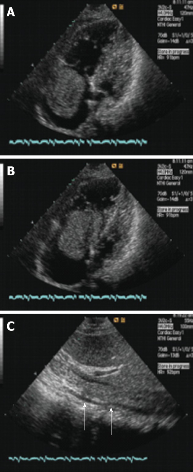
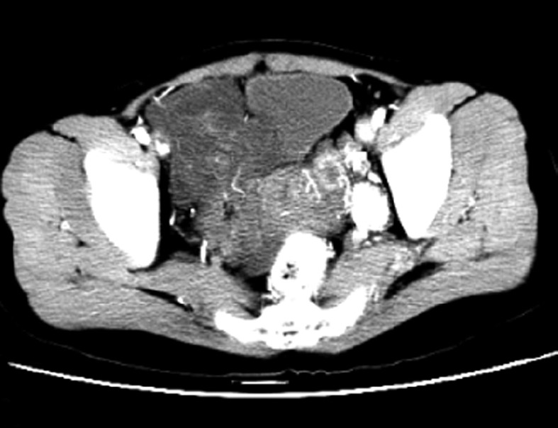
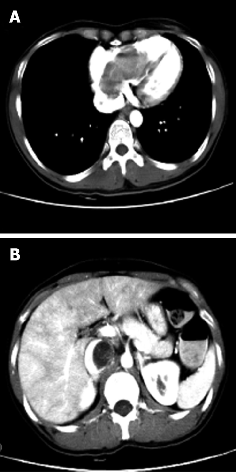
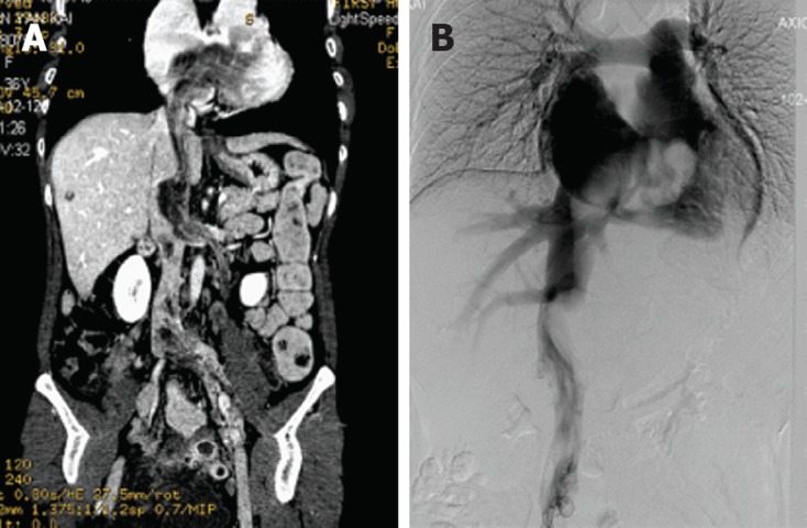
Intravenous leiomyomatosis (IVL), showing unusual growth patterns of uterine leiomyoma, is a rare neoplasm characterized by intravascular proliferation of a histologically benign-looking smooth muscle cell tumor mass, but not invading the tissue. To date, less than 300 cases have been reported and fewer than 100 cases with cardiac involvement. Imaging characteristics of IVL are still not clear so it is usually misdiagnosed before surgery. A 36-year-old woman, who had undergone hysterectomy due to hysteromyoma, presented with shortness of breath after activities. Imaging showed IVL with mass involvement of the left ovarian vein, left renal vein, left external and common iliac vein, as well as within the inferior vena cava (IVC), extending into the right atrium. The operation demonstrated that the mass had no stalk and had well-demarcated borders with the wall of the right atrium and IVC. The patient underwent a one-stage combined multidisciplinary thoraco-abdominal operation under general anesthetic. Subsequently, the pathology report confirmed IVL. IVL should be considered in a female patient presenting with an extensive mass in the right side of the heart. Imaging technology, such as echocardiogram, contrast-enhanced computed tomography and magnetic resonance imaging, can provide important information to reveal the mass, the range and path of the lesion, and relates to the surgical plan decision. Consequently, perfect and exact image examination is very necessary pre-operation.

Keywords: Computed tomography; Echocardiogram; Hysterectomy; Imaging; Intravenous leiomyomatosis lower.


Figures

Figure 1
Figure 1
Echocardiogram of the heart and inferior vena cava. A, B: Axial image demonstrates a mass within the right atrium that moves back and forth through the tricuspid orifice into the right ventricle in the systolic and diastolic period; C: Axial image demonstrates a mass within the inferior vena cava (arrows).
Figure 2
Figure 2
Preoperative pelvic contrast enhanced computed tomography shows a big irregular mass exhibiting heterogeneous enhancement.
Figure 3
Figure 3
Preoperative chest and abdominal computed tomography. A: Axial image demonstrates a lobulated mass within the right atrium and ventricle; B: Axial image shows a tumor in the inferior vena cava and resulting lumen stenosis.
Figure 4
Figure 4
Coronal plane computed tomography and digital subtraction angiography image of intravenous leiomyomatosis. A: Preoperative coronal plane computed tomography demonstrates a mass was present in the left ovarian vein, left renal vein, left external and common iliac vein, as well as within the inferior vena cava, extending into the right atrium; B: Digital subtraction angiography image demonstrates an irregular mass filling defect from the inferior vena cava to the right atrium.

References

    1. Birch-Hirschfeld FV. Lehrbuch der pathologischen Anatomie. 5th ed. Leipzig: FCW Vogel; 1896. p. 226.
    1. Baca López FM, Martínez-Enriquez A, Castrejón-Aivar FJ, Ruanova-León D, Yánez-Gutiérrez L. Echocardiographic study of an intravenous leiomyoma: case report and review of the literature. Echocardiography. 2003;20:723–725. doi: 10.1111/j.0742-2822.2003.02152.x). - DOI - PubMed
    1. Lee S, Kim DK, Narm KS, Cho SH. Pulmonary artery embolization of intravenous leiomyomatosis extending into the right atrium. Korean J Thorac Cardiovasc Surg. 2011;44:243–246. doi: 10.5090/kjtcs.2011.44.3.243). - DOI - PMC - PubMed
    1. Thian YL, Tan KH, Kwek JW, Wang J, Chern B, Yam KL. Leiomyomatosis peritonealis disseminata and subcutaneous myoma--a rare complication of laparoscopic myomectomy. Abdom Imaging. 2009;34:235–238. doi: 10.1007/s00261-008-9379-5. - DOI - PubMed
    1. Lee HJ, Choi J, Kim KR. Pulmonary benign metastasizing leiomyoma associated with intravenous leiomyomatosis of the uterus: clinical behavior and genomic changes supporting a transportation theory. Int J Gynecol Pathol. 2008;27:340–345. doi: 10.1097/PGP.0b013e3181656dab). - DOI - PubMed

LinkOut - more resources