Review
. 2012 Nov-Dec;1(6):861-78.
doi: 10.1002/wdev.79. Epub 2012 Jun 19.

The Caenorhabditis elegans epidermis as a model skin. I: development, patterning, and growth

Affiliations
Review

The Caenorhabditis elegans epidermis as a model skin. I: development, patterning, and growth

Andrew D Chisholm et al. Wiley Interdiscip Rev Dev Biol. 2012 Nov-Dec.

Abstract

The skin of the nematode Caenorhabditis elegans is composed of a simple epidermal epithelium and overlying cuticle. The skin encloses the animal and plays central roles in body morphology and physiology; its simplicity and accessibility make it a tractable genetic model for several aspects of skin biology. Epidermal precursors are specified by a hierarchy of transcriptional regulators. Epidermal cells form on the dorsal surface of the embryo and differentiate to form the epidermal primordium, which then spreads out in a process of epiboly to enclose internal tissues. Subsequent elongation of the embryo into a vermiform larva is driven by cell shape changes and cell fusions in the epidermis. Most epidermal cells fuse in mid-embryogenesis to form a small number of multinucleate syncytia. During mid-embryogenesis the epidermis also becomes intimately associated with underlying muscles, performing a tendon-like role in transmitting muscle force. Post-embryonic development of the epidermis involves growth by addition of new cells to the syncytia from stem cell-like epidermal seam cells and by an increase in cell size driven by endoreplication of the chromosomes in epidermal nuclei.


Figures

FIGURE 1
FIGURE 1. Anatomy, topology, and genealogy of the epidermis
(A) Anatomy of the C. elegans epidermis in L1 stage. Lateral views showing nuclei and cell boundaries, based on Sulston et al. (1983) and WormAtlas. In this and other figures, hyp7 is green, seam cells (H, V, T) are dark blue, ventral epidermal (P) cells are red; hyp4 is pink, hyp5 light blue, and hyp6 teal. (B) Cylindrical projections of the epidermis, separated at ventral midline and unrolled so that right is up and anterior to the left. The dorsal midline is indicated. Projections of the initial embryonic epithelium prior to cell fusion, L1 stage epidermis, and adult. The numbers and approximate disposition of cells are correct; the exact pattern of cell contacts is simplified. For more anatomically accurate projections, see Wormbook hypFIG2.
FIGURE 2
FIGURE 2. Lineage origins of the embryonic and post-embryonic epidermis
(A) Abbreviated embryonic cell lineage showing the origin of epidermal precursors from the AB and C lineages . Cells are named according to standard C. elegans lineage nomenclature: AB and C are early embryonic blastomeres; a/p indicates anterior or posterior daughter and l/r indicates left or right daughter. Epidermal potential is intrinsic to AB and C and segregates at asymmetric divisions at the AB and C stages (i.e., at the division of ABar to ABara and ABarp, and so on) to five major epidermal precursors (filled circles). Epidermal cells are born in two successive rounds of cell division. 66 major epidermal cells (hyp4–7, seam, and P cells, shown in the projections) are born at ~240 minutes and comprise the initial epidermal primordium that forms dorsally and encloses the embryo. The remaining 12 minor epidermal cells of the head and tail (hyp1–3 and 8–11) are born in the next round of AB divisions (270–300 min) from ‘minor epidermal precursors’ (open circles) and are not shown in the cylindrical projections; 16 other epidermal-like cells (arcade, XXX, rectal epithelial cells, tail spike) are also born in this round of divisions and are not shown here. Numbers under cell names indicate the number of major and minor (gray) cells derived from each precursor. Projections show only the 66 major epidermal cells; note that except for V3 and V5, lineally related cells are adjacent in the epithelium. Color code as in FIGURE 1. (B) Representative lineage of a post-embryonic epidermal seam cell such as V1, illustrating asymmetric divisions in each larval stage and the L2-specific doubling division.
FIGURE 3
FIGURE 3. Transcriptional hierarchy regulating epidermal identity and differentiation
Regulatory hierarchy in embryonic epidermal specification, based on , and . Lineage-specific regulators activate the ‘epidermal organ identity’ gene ELT-1 in descendants of the AB and C blastomeres. ELT-1 initiates epidermal identity by inhibiting other organ identity modules and by directly activating three epidermal ‘differentiation’ factors: LIN-26, ELT-1, and NHR-25. LIN-26 confers generic epithelial characteristics . ELT-3 promotes epidermal-specific aspects of terminal differentiation (e.g. cuticle collagen expression) but is not itself essential for epidermal development. The three differentiation factors appear to indirectly repress one another via negative feedback on ELT-1. NHR-23 and Grainyhead/GRH-1 promote other aspects of epidermal differentiation, but their location in the hierarchy has not been defined. In parallel, lineage-specific regulators specify subtypes of epidermal cell. Dorsal epidermal cells require the T-box genes TBX-8, 9; in C-derived hyp7, PAL-1 likely activates TBX-8/9 directly. One target of TBX-8/9 in C-derived hyp7 is the even-skipped ortholog VAB-7 . DIE-1 is expressed in dorsal hyp7 in response to unknown signals. Lateral epidermal cells are specified by CEH-16 and ELT-5/6, which in turn repress the differentiation factor ELT-3. Post-embryonic seam cells are maintained in a stem-cell like state by the action of RNT-1/Runx and BRO-1/CBFβ; BRO-1 is directly activated by ELT-1 . Dorsal and ventral cells (all non-seam epidermis) express ELT-3.
FIGURE 4
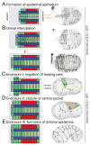
FIGURE 4. Embryonic epidermal morphogenesis
Early stages of epidermal morphogenesis, illustrated as projections and in frames from movies of the epidermal junctional marker DLG-1::GFP (xnIs17). (A) Initial formation of the dorsal epidermal epithelium, ~230–250 minutes post first cleavage. The epidermis occupies the posterior 2/3 of the dorsal part of the embryo. (B) Dorsal intercalation (~250–390 minutes), early and late stages. Intercalation is not as synchronous or as consistent as in the cartoon. (C) Ventral enclosure: migrations of four leading cells to the ventral midline, 365–375 min. (D) Ventral enclosure II: closure of the ventral pocket by P cells and hyp7 ventral cells, 370–385 min. (E) Enclosure III: formation of the anterior epidermis, 380–395 min. The leading cells move anteriorly; hyp4 and hyp5 cells enclose the head in short-range movements. Dorsal hyp7 cells begin to fuse. DLG-1::GFP images in A, B are dorsal views, from Movie 2 in Chisholm and Hardin 2005, WormBook. Frames in C-E are ventral views (C.A. Giurumescu and A.D.C., unpublished).
FIGURE 5
FIGURE 5. Anteroposterior pattern in the larval epidermis
Patterning of the seam in the trunk and tail is specified by genes in the C. elegans Hox gene cluster. Schematic showing Hox cluster genes on part of chromosome III; putative Drosophila homologs are indicated . Examples of cell fate transformations: in mab-5 mutants the posterior seam cells V5 and V6 adopt V1–V4-like fates (‘V4’) . Patterning of the head seam cells is specified by the PAX-6 locus vab-3/mab-18. , . In pax-6 null mutants the anterior-most seam cells H0 and H1 are transformed to H2-like fates. Hox cluster genes also regulate anteroposterior pattern in the ventral epidermal (P) cells.

References

    1. Fuchs E. Scratching the surface of skin development. Nature. 2007;445:834–842. - PMC - PubMed
    1. Thompson DP, Geary TG. Excretion/Secretion, Ionic and Osmotic Regulation. In: Lee DL, editor. The Biology of Nematodes. Boca Raton, FL: CRC Press; 2002. pp. 291–320.
    1. Payre F. Genetic control of epidermis differentiation in Drosophila. Int J Dev Biol. 2004;48:207–215. - PubMed
    1. Le Guellec D, Morvan-Dubois G, Sire JY. Skin development in bony fish with particular emphasis on collagen deposition in the dermis of the zebrafish (Danio rerio) The International journal of developmental biology. 2004;48:217–231. - PubMed
    1. Koster MI, Roop DR. Mechanisms regulating epithelial stratification. Ann Rev Cell Dev Biol. 2007;23:93–113. - PubMed

Publication types

Substances

LinkOut - more resources