. 2013 Jun 15;73(12):3725-36.
doi: 10.1158/0008-5472.CAN-12-3468. Epub 2013 Mar 28.

Androgen receptor-independent function of FoxA1 in prostate cancer metastasis

Affiliations

Androgen receptor-independent function of FoxA1 in prostate cancer metastasis

Hong-Jian Jin et al. Cancer Res. .

Abstract

FoxA1 (FOXA1) is a pioneering transcription factor of the androgen receptor (AR) that is indispensible for the lineage-specific gene expression of the prostate. To date, there have been conflicting reports on the role of FoxA1 in prostate cancer progression and prognosis. With recent discoveries of recurrent FoxA1 mutations in human prostate tumors, comprehensive understanding of FoxA1 function has become very important. Here, through genomic analysis, we reveal that FoxA1 regulates two distinct oncogenic processes via disparate mechanisms. FoxA1 induces cell growth requiring the AR pathway. On the other hand, FoxA1 inhibits cell motility and epithelial-to-mesenchymal transition (EMT) through AR-independent mechanism directly opposing the action of AR signaling. Using orthotopic mouse models, we further show that FoxA1 inhibits prostate tumor metastasis in vivo. Concordant with these contradictory effects on tumor progression, FoxA1 expression is slightly upregulated in localized prostate cancer wherein cell proliferation is the main feature, but is remarkably downregulated when the disease progresses to metastatic stage for which cell motility and EMT are essential. Importantly, recently identified FoxA1 mutants have drastically attenuated ability in suppressing cell motility. Taken together, our findings illustrate an AR-independent function of FoxA1 as a metastasis inhibitor and provide a mechanism by which recurrent FoxA1 mutations contribute to prostate cancer progression.


Conflict of interest statement

Disclosure of Potential Conflicts of Interest

No potential conflicts of interest were disclosed.

Figures

Figure 1
Figure 1. Expression microarray analyses reveal distinct roles of FoxA1 in inducing cell growth but inhibiting cell motility
A, Western blot of FoxA1 protein in control and knockdown LNCaP cells. GAPDH was used as a loading control. B–C, Heatmap view and GO analysis of FoxA1-induced (B) and -repressed genes (C). Differentially expressed genes were identified through microarray analysis of control and FoxA1-knockdown LNCaP cells using a 2-fold cutoff and p-value threshold of 0.01. GO concepts involved in cell cycle are shown in red bars, cell motility in green and androgen response in blue. The P values and the number of genes in each GO category are indicated at the x-axis and next to the bar, respectively. D–E, LNCaP shFoxA1 and control cells were cultured in hormone-depleted medium or treated with 1 nM R1881 for 24 hours. Total RNA was isolated and analyzed by microarrays. Expression levels of FoxA1-induced cell cycles genes (D) and FoxA1-repressed cell migration genes (E) were determined.
Figure 2
Figure 2. FoxA1 positively regulates cell proliferation requiring the AR
A–B, WST-1 cell growth assay of AR-positive LNCaP (A) and 22RV1 (B) control (shCtrl) and shRNA-treated (shFoxA1) cells. Data shown are the mean of independent replicates +/− SEM. FoxA1 protein knockdown was confirmed by western blot (inset). C–D, AR-negative PC-3M (C) and DU145 (D) cells were subjected to control and FoxA1 overexpression. Total protein was blotted (inset) and cell growth assayed. Data shown are the mean of independent replicates +/− SEM. E–F, PC-3M cells were transfected with control, FoxA1- or AR-expressing vector, or both and stable cell lines were established. AR and FoxA1 protein were determined by immunoblotting (E) and cell growth by WST-1 assay (F). G, Control (shCtrl) and FoxA1-knockdown (shFoxA1) LNCaP cells were treated with vehicle or 10nM anti-androgen bicalutamide. Total cell growth was measured by WST-1 assay.
Figure 3
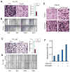
Figure 3. FoxA1 inhibits PCa cell invasion and migration independent of AR
A–D, Ectopic FoxA1 overexpression decreased DU145 and PC-3M cell invasion (A, C) and migration (B, D). DU145 and PC-3M cells were infected with control and FoxA1-expressing lentivirus and subjected to antibiotic selection. Stable control and FoxA1-expressing cells were then analyzed using Boyden Chamber invasion assay for 24 hours. Invaded cells were quantified using colormetry with absorbance at 560nm (right panels of A and C). Cell migration was measured using wound-healing assay at a time-course after cell scratching. E–F, FoxA1 knockdown increases LNCaP cell invasion. Control and FoxA1-knockdown cells were established and grown in hormone-deprived or androgen-treated medium and assessed by Boyden Chamber Assay. Error Bars indicate the mean from three independent experiments +/− SEM.
Figure 4
Figure 4. FoxA1 suppresses Epithelial-to-Mesenchymal Transition
A, Western blot analysis of E-Cad, FoxA1 and VIM in a panel of PCa cell lines. B, Phase-contrast microscopy images of control vs. FoxA1-knockdown LNCaP cells. Scale bars represent 50 μm. C, qRT-PCR showing increased VIM expression in FoxA1-knockdown LNCaP cells. D. Western blot of VIM protein following FoxA1 knockdown. LNCaP cell lysates were first immunoprecipitated with a mouse anti-VIM antibody and then detected using a rabbit anti-VIM antibody. Total cell lysate (bottom) was immunoblotted by anti-GAPDH as a loading control. E, Phase-contrast microscopy images of control vs. PC-3M cells with ectopic FoxA1 overexpression. Scale bars represent 50 μm. F–G, Induced levels of E-Cad transcript and protein in FoxA1-expressing PC-3M cells.
Figure 5
Figure 5. SLUG is a direct target of FoxA1 and a key mediator of its anti-motility function
A, FoxA1 ChIP-Seq in the control and FoxA1-knockdown LNCaP cells around the SLUG gene. A major binding event locates at a downstream enhancer within the intragenic region, around which the primers SLUG pF1 and pR1 were designed for ChIP-PCR in B-C and Supplementary Figure S5A. B. FoxA1 ChIP-PCR in LNCaP cells. IgG was used as a control antibody and PSA as a positive control gene. C. FoxA1 binding was validated by ChIP-PCR in control and FoxA1-knockdown LNCaP cell. D, LNCaP, VCaP and 22RV1 cells with stable FoxA1 knockdown were established. SLUG gene expression was assessed by qRT-PCR. E, qRT-PCR analysis of SLUG gene expression in LNCaP, 22RV1, VCaP, RWPE and DU145 cells with control or FoxA1 overexpression. F, LNCaP cells were transfected with control- or FoxA1-targeting shRNA lentivirus and selected for stable cells. Cell lysates were collected over a 7-day period and total protein was immunoblotted. G, Protein levels of FoxA1 and SLUG in control vs. FoxA1-expressing RWPE cells. H, qRT-PCR analysis of SLUG gene expression in control and FoxA1-knockdown LNCaP cells grown in the presence or absence of androgen. I–J, SLUG knockdown reversed shFoxA1-induced cell invasion. Cell invasion was assessed by Boyden Chamber Assay (I) and quantified using colorimetry (absorbance at 560nm) (J).
Figure 6
Figure 6. FoxA1 inhibits xenograft prostate tumor metastasis in mice
A–C, FoxA1 overexpression inhibits orthotopic tumor growth and metastasis in mice. Nude mice were injected orthotopically with luciferase-labeled control and FoxA1-expressing PC-3M cells. Xenogen images of two representative mice per group are shown in A. Tumor growth (B) and metastasis (C) were monitored through living mice imaging using an IVIS Spectrum Bioluminescence System. D, FoxA1 mRNA level in a microarray dataset profiling 33 benign prostate, 81 localized prostate tumor and 40 metastatic PCa (22). Significant P-values are shown. E, qRT-PCR analysis of FoxA1 mRNA in a panel of human prostate tissues. Significant P-value was shown.
Figure 7
Figure 7. Mutant FoxA1 failed to inhibit SLUG expression and cell invasion
A–B, Wild type (WT) FoxA1, FoxA1 F400I and FoxA1 M253K were stably expressed in DU145 cells. Stable cells were generated using a different lentivirus construct or empty vector (as a control). QRT-PCR(A) and western blotting (B) confirmed FoxA1 expression. C, Wild type FoxA1 overexpression significantly inhibited SLUG expression. FoxA1 mutations resulted in significantly recovered SLUG expression. D–E, Cell invasion was assessed by Boyden Chamber Assay (D) and quantified using colorimetry (absorbance at 560nm) (E).

References

    1. Lee CS, Friedman JR, Fulmer JT, Kaestner KH. The initiation of liver development is dependent on Foxa transcription factors. Nature. 2005;435:944–7. - PubMed
    1. Gao N, LeLay J, Vatamaniuk MZ, Rieck S, Friedman JR, Kaestner KH. Dynamic regulation of Pdx1 enhancers by Foxa1 and Foxa2 is essential for pancreas development. Genes & development. 2008;22:3435–48. - PMC - PubMed
    1. Bernardo GM, Lozada KL, Miedler JD, Harburg G, Hewitt SC, Mosley JD, et al. FOXA1 is an essential determinant of ERalpha expression and mammary ductal morphogenesis. Development. 2010;137:2045–54. - PMC - PubMed
    1. Kaestner KH. The FoxA factors in organogenesis and differentiation. Current opinion in genetics & development. 2010;20:527–32. - PMC - PubMed
    1. Sekiya S, Suzuki A. Direct conversion of mouse fibroblasts to hepatocyte-like cells by defined factors. Nature. 2011;475:390–3. - PubMed

Publication types

MeSH terms

Substances

Associated data