Review
. 2013 Apr;131(4):933-57; quiz 958.
doi: 10.1016/j.jaci.2013.02.023.

B-lymphocyte lineage cells and the respiratory system

Affiliations
Review

B-lymphocyte lineage cells and the respiratory system

Atsushi Kato et al. J Allergy Clin Immunol. 2013 Apr.

Abstract

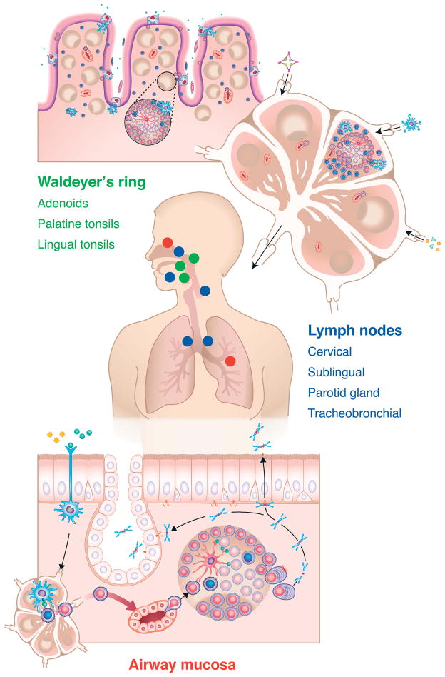
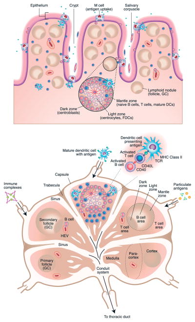
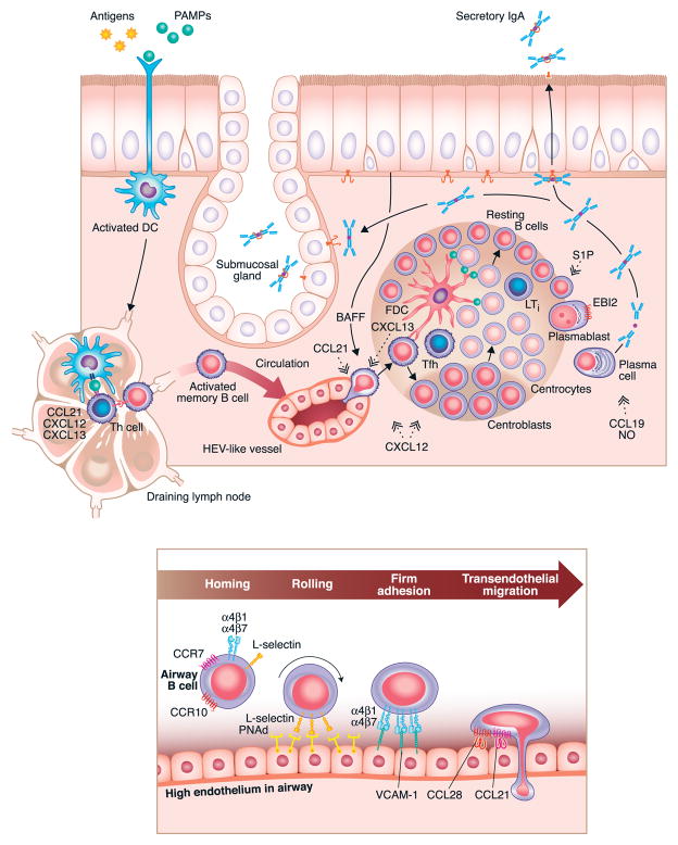
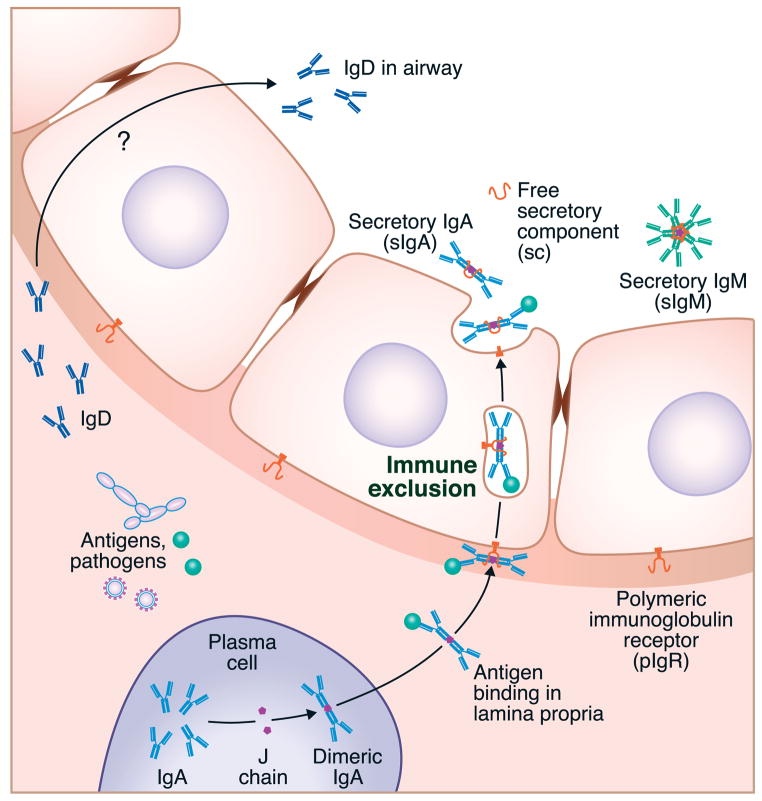
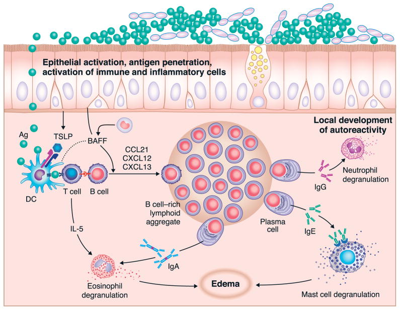
Adaptive humoral immune responses in the airways are mediated by B cells and plasma cells that express highly evolved and specific receptors and produce immunoglobulins of most isotypes. In some cases, such as autoimmune diseases or inflammatory diseases caused by excessive exposure to foreign antigens, these same immune cells can cause disease by virtue of overly vigorous responses. This review discusses the generation, differentiation, signaling, activation, and recruitment pathways of B cells and plasma cells, with special emphasis on unique characteristics of subsets of these cells functioning within the respiratory system. The primary sensitization events that generate B cells responsible for effector responses throughout the airways usually occur in the upper airways, tonsils, and adenoid structures that make up the Waldeyer ring. On secondary exposure to antigen in the airways, antigen-processing dendritic cells migrate into secondary lymphoid organs, such as lymph nodes, that drain the upper and lower airways, and further B-cell expansion takes place at those sites. Antigen exposure in the upper or lower airways can also drive expansion of B-lineage cells in the airway mucosal tissue and lead to the formation of inducible lymphoid follicles or aggregates that can mediate local immunity or disease.


Figures

Figure 1
Figure 1
Overview of the organization of tissues involved in the induction, elicitation and effector responses of B cells and plasma cells in the airways of humans. Shown in green text are the components of Waldeyer’s ring, including the adenoid(s) and palatine and lingual tonsils (shown in green dots on the transverse model of a human). The blue text and blue dots in the model indicate lymph nodes that are centrally involved in sensitization and elicitation of airways responses. These include cervical, sublingual and parotid nodes, as well as tracheobronchial nodes. The figure at the bottom displays an effector response in the airways (for details, see Figure 3A), which could occur either in the lower airways or the upper airways and sinuses, as indicated by the red dots in the transverse section of the model. An important point is that the primary sensitization events that generate B cells responsible for effector responses throughout the airways often occur in the upper airways. For further discussion, please see the main text of the review.
Figure 2
Figure 2
Detailed overview of primary inductive lymphoid tissues contained in tonsils (2A – top) and lymph nodes (2B – bottom). Tonsils and adenoid tissue contain M cells that mediate antigen uptake into the tissue that is rich with lymphoid follicles in which the primary expansion of naïve B cells occurs, followed by the subsequent generation of memory B cells that populate other lymphoid tissues, especially lymph nodes. Dark and light zones, containing centroblasts and centrocytes, are shown, and the mantle zone, in which dendritic cells, B cells and T cells collaborate for B cell activation, are illustrated in the magnified inset. A similar expansion of memory (and naïve) B cells can occur with secondary exposure in the lymph nodes that are draining the airways (2B). Detailed discussion of the cellular dynamics in these primary and secondary expansions of antigen specific B cells is found within the text.
Figure 3
Figure 3
Representative effector response within airway tissue (3A – top) in which antigen exposure leads to local recruitment of memory (and perhaps naïve) B cells (3B – bottom) and subsequent local expansion of B cells and plasma cells within the airway tissue. The chemokines that drive the recruitment of B cells from the circulation include CCL21, and those involved in localization within quasi-organized inducible lymphoid tissue include CXCL12 and CXCL13 as well as CCL19 and CCL21. The expansion of B cells within the tissue bears a resemblance to the initial induction and expansion of B cells discussed in Figure 1 and in the text. Recruitment of circulating B cells occurs partly via transendothelial migration across endothelium that resembles HEV found in lymph nodes. Some of the molecular participants in the rolling, firm adhesion and transendothelial migration of B cells into the tissue are described in Figure 3B.
Figure 4
Figure 4
The principle of immune exclusion. Local expression of immunoglobulins of the IgA and IgM isotype can be leveraged for the removal of antigens, allergens and pathogens via the polymeric immunoglobulin receptor (PIgR). PIgR binds to multimeric forms of IgA and IgM containing the J chain and moves them across the epithelium by a vesicular transport mechanism that is robust enough to transport whole organisms bound to the immunoglobulin.
Figure 5
Figure 5
Overview of the role of B cells in chronic rhinosinusitis. Studies of the pathogenesis of the polypoid form of CRS have implicated B cells and several isotypes of immunoglobulins in the pathogenesis of this important and common disease. Elevated levels of B cells and plasma cells are established in CRS and are accompanied by elevated levels of the chemokines that attract them (including CXCL12 and CXCL13) as well as TSLP and BAFF. Formation of organized follicular structures occurs in some cases of CRS. Although the specificity of the IgG, IgA and IgE antibodies made in nasal polyp tissue is not completely known, recent evidence shows the presence of autoantibodies of the IgA and IgG isotypes. Cellular targets of immunoglobulins that are enriched in nasal polyps include eosinophils (which respond briskly to IgA activation), mast cells (which respond to IgE receptor crosslinking) and neutrophils (which respond to IgG immune complexes). Release of mediators by these cell types is likely to be important in the pathogenesis of the disease, including formation of nasal polyps.

References

    1. Salvi S, Holgate ST. Could the airway epithelium play an important role in mucosal immunoglobulin A production? Clin Exp Allergy. 1999;29:1597–605. - PubMed
    1. Drolet JP, Frangie H, Guay J, Hajoui O, Hamid Q, Mazer BD. B lymphocytes in inflammatory airway diseases. Clin Exp Allergy. 2010;40:841–9. - PubMed
    1. Wilkes DS, Twigg HL., 3rd B-lymphocytes in the lung: a topic to be revisited. Sarcoidosis Vasc Diffuse Lung Dis. 2001;18:34–49. - PubMed
    1. Brandtzaeg P. Potential of nasopharynx-associated lymphoid tissue for vaccine responses in the airways. Am J Respir Crit Care Med. 2011;183:1595–604. - PubMed
    1. Kurosaki T, Shinohara H, Baba Y. B cell signaling and fate decision. Annu Rev Immunol. 2010;28:21–55. - PubMed

Publication types

MeSH terms