. 2013 Mar 28;153(1):126-38.
doi: 10.1016/j.cell.2013.03.018.

Somatic mutations of the immunoglobulin framework are generally required for broad and potent HIV-1 neutralization

Affiliations

Somatic mutations of the immunoglobulin framework are generally required for broad and potent HIV-1 neutralization

Florian Klein et al. Cell. .

Abstract

Broadly neutralizing antibodies (bNAbs) to HIV-1 can prevent infection and are therefore of great importance for HIV-1 vaccine design. Notably, bNAbs are highly somatically mutated and generated by a fraction of HIV-1-infected individuals several years after infection. Antibodies typically accumulate mutations in the complementarity determining region (CDR) loops, which usually contact the antigen. The CDR loops are scaffolded by canonical framework regions (FWRs) that are both resistant to and less tolerant of mutations. Here, we report that in contrast to most antibodies, including those with limited HIV-1 neutralizing activity, most bNAbs require somatic mutations in their FWRs. Structural and functional analyses reveal that somatic mutations in FWR residues enhance breadth and potency by providing increased flexibility and/or direct antigen contact. Thus, in bNAbs, FWRs play an essential role beyond scaffolding the CDR loops and their unusual contribution to potency and breadth should be considered in HIV-1 vaccine design.


Figures

Figure 1
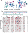
Figure 1. Somatic mutations in the framework regions of HIV-1-reactive antibodies
(A) Ribbon representation of the variable domains of 3BNC60 (Scheid et al., 2011), illustrating the CDRs (magenta) and the FWRs of the immunoglobulin heavy (blue) and light (cyan) chain. (B) Illustration of Kabat and IMGT CDR (magenta) and FWR (IgH – blue; IgL – cyan) assignments for the variable heavy and light chain domains of 3BNC60. Gray arrows indicate β-strands defined by the crystal structure of the 3BNC60 Fab (Scheid et al., 2011). (C) Position of FWR mutations in heavy and light chain of the 17 investigated antibodies with broad neutralizing activity (see also Supplementary Data 1). Indicated are silent (black) and replacement (red) mutations. Insertions are illustrated in blue. Number of replacement mutations within CDR1/2 and FWR1–4 are listed in the two columns at the very right (see also Table S1). HIV-1 reactive antibodies with limited neutralization are displayed in Figure S2.
Figure 2
Figure 2. Binding and neutralization activity of mature and FWR-reverted (FWR-GL) broad neutralizing antibodies
Evaluation of binding to gpl40YU2 ELISA (left) of mature antibodies (green) and antibodies with FWRs reverted to germline (FWR-GL; blue, Supplementary Data 1B). Panels on the right compare IC50 values for neutralization of Tier 1 (MW965.26, SF162.LS, Bal.26, SS1196.1, DJ263.8, 6535.3) Tier 2 (RHPA4259.7, SC422661.8, TRO.11, YU2.DG), and Tier 3 (PV0.4) viruses (Table S2). Only viruses are shown that were neutralized by the mature version of the antibody. Neutralization activity is color-coded (blue arrow: < 0.001 µg/ml; dark red: IC50 between 0.001 and 0.01 µg/ml; red: > 0.01 – 0.1 µg/ml; orange: > 0.1 – 1 µg/ml; light orange: > 1 – 10 µg/ml; yellow: > 10 µg/ml; white: IC50 was not achieved up to the tested concentration). Antibodies (NIH45–46, 3BN60) with reverted CDR1/2 are shown in Figure S2A and HIV-1 reactive antibodies with limited neutralization are displayed in Figure S2B. The FWRs of VRC01, 3BNC60 and 3BNC131 were also reverted according to IMGT (Giudicelli et al., 2006) and results are shown in Figure S2C. A detailed illustration of the FWR mutations in 2G12 is shown in Figure S3.
Figure 3
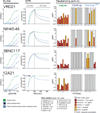
Figure 3. Binding and neutralization activity of mature and FWR-reverted (FWR-GL and FWR-GLCR+) broad neutralizing antibodies
Evaluation of binding to gpl40YU2 ELISA (left) and SPR (middle) of mature antibodies (green), antibodies with FWRs reverted to germline (FWR-GL, blue), and antibodies with germline-reverted FWRs except gp120-contacting residues (FWR-GLCR+; light blue; Supplementary Data ID). SPR results are shown for starting concentrations of 1 µM. BD (below detection; no binding of the antibody was observed). Panels on the right compare IC50 values for neutralization of Tier 1 to Tier 3 viruses (Table S2) as in Figure 2. Neutralization activity is color-coded as indicated.
Figure 4
Figure 4. Effects of a FWR insertion and a C” β-strand proline in clone RU01
Phylogenetic tree of the antibodies (Ig heavy chain) derived from the RU01 clone that members include 3BNC117 and 3BNC60 (Scheid et al., 2011). Antibodies that carry both the 4 amino acid insertion in FWR3 and the A61P somatic mutation are shown in red, antibodies with only the A61P mutation are shown in orange, and antibodies without either feature are shown in black. Bootstrap values (1000 trials, seed=111) of the phylogenetic tree are indicated. Structure of 3BNC117 IGVH in its gpl20-bound conformation is shown in Figure S5.
Figure 5
Figure 5. Analysis of 3BNC60 insertion
(A) Superimposition of the structure of the VH domain of 3BNC60 (cyan) (Scheid et al., 2011) onto the NIH45–46 VH domain from cocrystal structure of NIH45–46 (gray) bound to gpl20 (gold) (Diskin et al., 2011) highlighting the 4 residue insertion in FWR3 of 3BNC60 (cyan arrow) and a potential interaction between the insertion and the gpl20 V1/V2 loop (note that the V1/V2 loop was truncated in the gpl20 construct used for co-crystallization with NIH45–46 and VRC01) (Diskin et al., 2011; Zhou et al., 2010). (B) Alignment of the heavy chain FWR3 sequences of 3BNC60 (Scheid et al., 2011), 3BNC60 without the 3BNC60 insertion (3BNC60∆I), 3BNC55 (Scheid et al., 2011) and 3BNC55 containing the 3BNC60 insertion (3BNC55+I; Supplementary Data 1E). The 4 amino acid insertion in FWR3 of 3BNC60 is shown in cyan and the region grafted into 3BNC60 from 3BNC55 in order to delete the insertion without disrupting the structure is shown in gray. The amino acid changes to introduce the insertion into 3BNC55 are shown in cyan. (C) In vitro neutralization data (IC50 values in µg/ml) comparing the potencies of the antibodies. The upper panel compares neutralization of selected viral strains by 3BNC60 and, 3BNC60∆I. The lower panel compares the neutralization by a less potent member of the RU01 clone (3BNC55), in which the FWR3 insertion of 3BNC60 was introduced (3BNC55+I). Reduced and increased neutralization activity of the engineered antibodies (3BNC60∆I, 3BNC55+I) is highlighted red and green, respectively. Neutralization data on VRC01 with and without the 3BNC60 insertion is displayed in Table S2.
Figure 6
Figure 6. Comparison of 3BNC60 and 3BNC60P61A structures, thermal denaturation profiles and neutralizing activities
(A, left) 3BNC60P61A (magenta heavy, chain; yellow, light chain) was superimposed on the structure of 3BNC60 (cyan, heavy chain; red, light chain; Table S3). The C” β-strand within FWR3 of the VH domain differs in conformation between the two structures. (A, middle/right) Close-up of the C’ and C” β-strands of 3BNC60P61A (magenta), 3BNC117 bound to gol20 (yellow), 3BNC60 (cyan), and a murine Fab (green). The main chain atoms of the VH domain C’ and C” β-strands of 3BNC60P61A exhibit a typical hydrogen bonding pattern for an anti-parallel β-sheet. Three of the five inter-β-strand hydrogen bonds in 3BNC60P61A are found in all three structures (yellow dashed lines), whereas 3BNC60 lacks two and the murine Fab/3BNC117 lacks one of the hydrogen bonds (green dashed lines in 3BNC60P61A). Co-crystallization of the 3BNC60-relative 3BNC117 with gpl20 shows that Proline61 is accommodated without disrupting the C' - C" β-sheet when 3BNC117 is bound to gpl20. Overview of the packing in crystals of 3BNC60 and 3BNC60P61A is shown in Figure S6. (B) Thermal denaturation profiles of the 3BNC60 and 3BNC60P61A Fabs monitored by the CD signal at 218 nm. TmS (indicated with arrows) were derived by estimating the halfpoint of the ellipticity change between the beginning and end of each transition. (C) In vitro neutralization data (IC50 values in µg/ml) comparing the 3BNC60 and 3BNC60P61A IgGs for a panel of 19 viruses. Reduced neutralization activity is highlighted in red. Additional 9 viral strains (T250-4, T278-50, 620345.C01, X2088_c9, 89-Fl_2_25, 6540.v4.c1, CAP45.2.00.G3, 6545.v4.c1, Du422.1) were resistant to both 3BNC60 and 3BNC60P61A. The 3BNC60P61A mutant was not significantly more potent than 3BNC60 against any of the 27 strains tested.

References

    1. Adams PD, Afonine PV, Bunkoczi G, Chen VB, Davis IW, Echols N, Headd JJ, Hung LW, Kapral GJ, Grosse-Kunstleve RW, et al. PHENIX: a comprehensive Python-based system for macromolecular structure solution. Acta Crystallogr D Biol Crystallogr. 2010;66:213–221. - PMC - PubMed
    1. Amit AG, Mariuzza RA, Phillips SE, Poljak RJ. Three-dimensional structure of an antigen-antibody complex at 2.8 A resolution. Science. 1986;233:747–753. - PubMed
    1. Amzel LM, Poljak RJ. Three-dimensional structure of immunoglobulins. Annu Rev Biochem. 1979;48:961–997. - PubMed
    1. Batista FD, Neuberger MS. Affinity dependence of the B cell response to antigen: a threshold, a ceiling, and the importance of off-rate. Immunity. 1998;8:751–759. - PubMed
    1. Buchacher A, Predl R, Strutzenberger K, Steinfellner W, Trkola A, Purtscher M, Gruber G, Tauer C, Steindl F, Jungbauer A, et al. Generation of human monoclonal antibodies against HIV-1 proteins; electrofusion and Epstein-Barr virus transformation for peripheral blood lymphocyte immortalization. AIDS research and human retroviruses. 1994;10:359–369. - PubMed

Publication types