Review
. 2013 May-Jun;58(3):203-32.
doi: 10.1016/j.survophthal.2012.08.008. Epub 2013 Mar 27.

Serpiginous choroiditis and infectious multifocal serpiginoid choroiditis

Affiliations
Review

Serpiginous choroiditis and infectious multifocal serpiginoid choroiditis

Hossein Nazari Khanamiri et al. Surv Ophthalmol. 2013 May-Jun.

Abstract

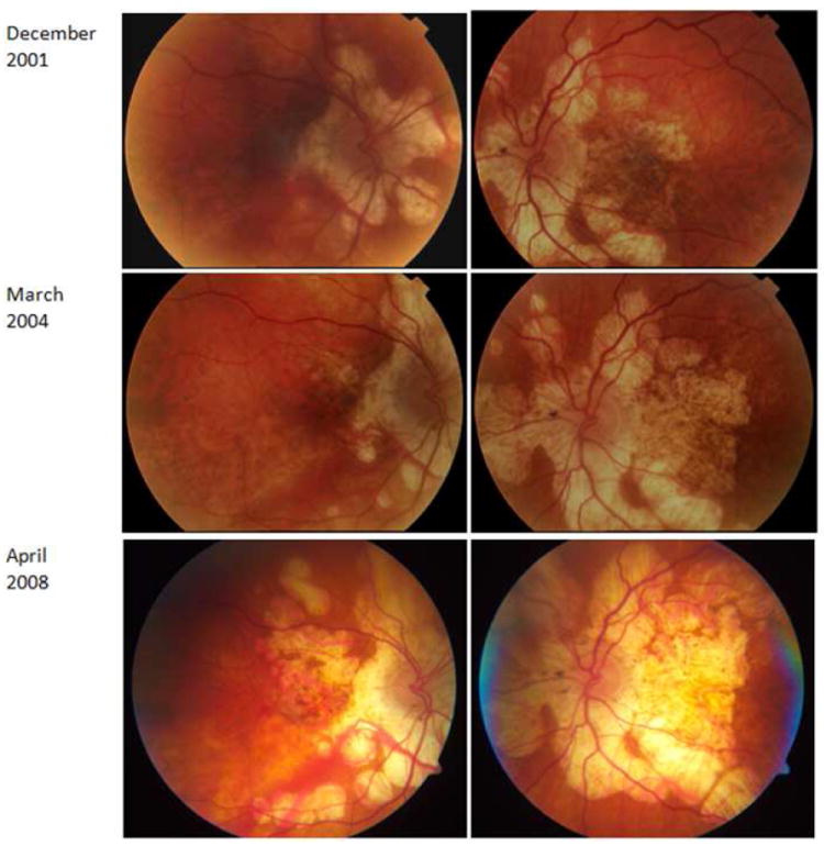
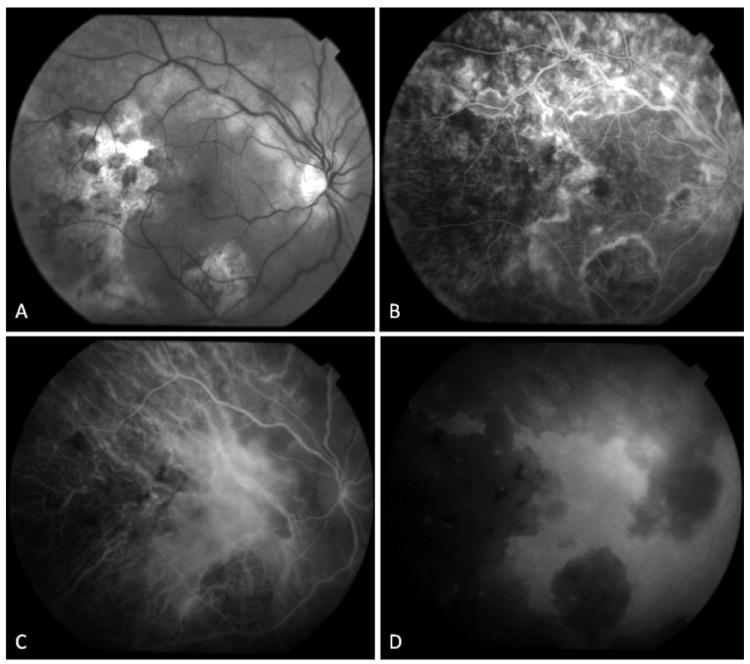
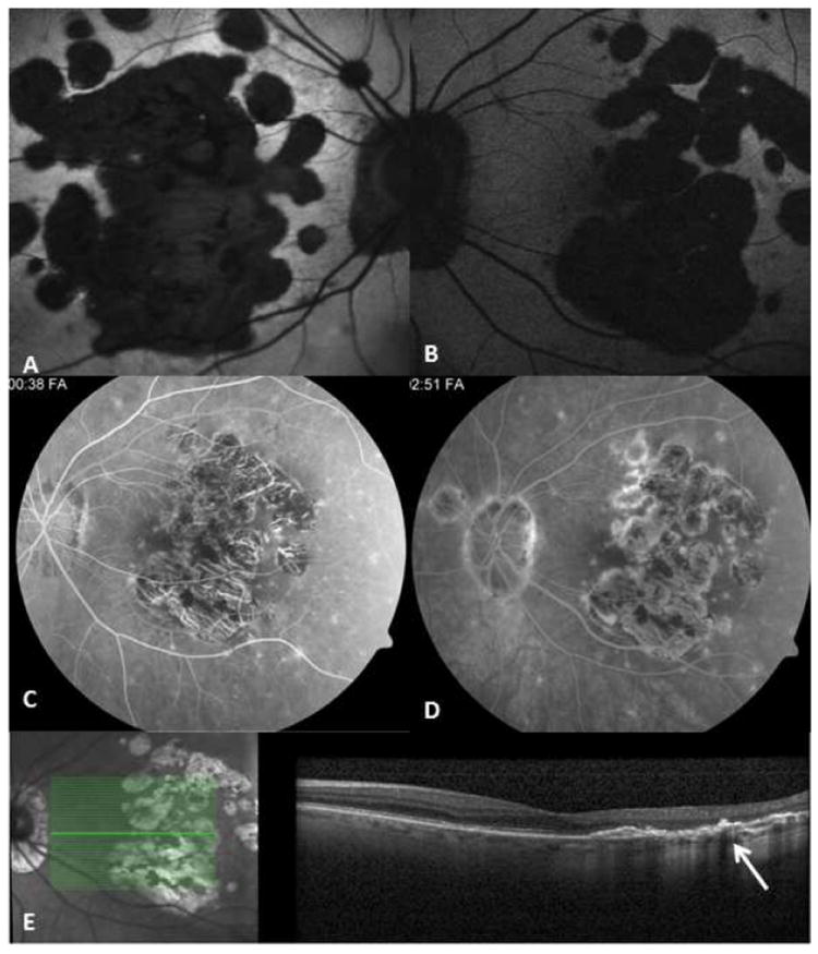
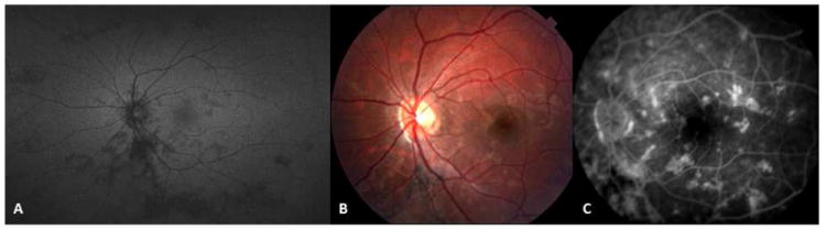
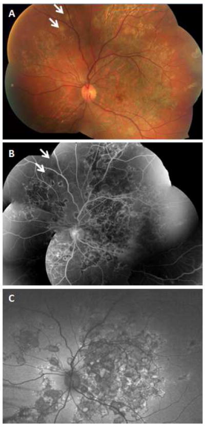
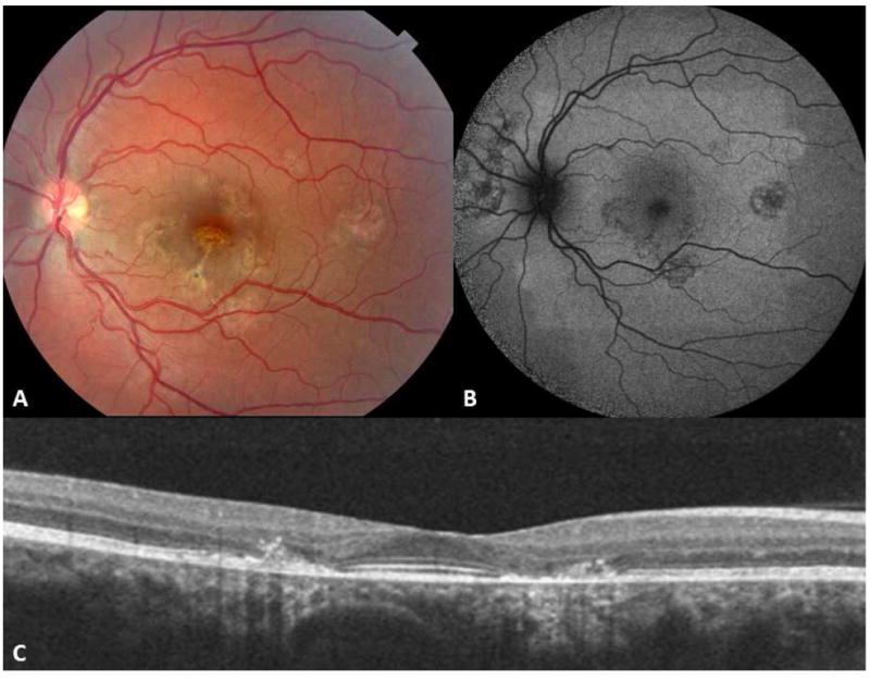
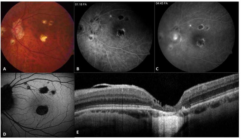
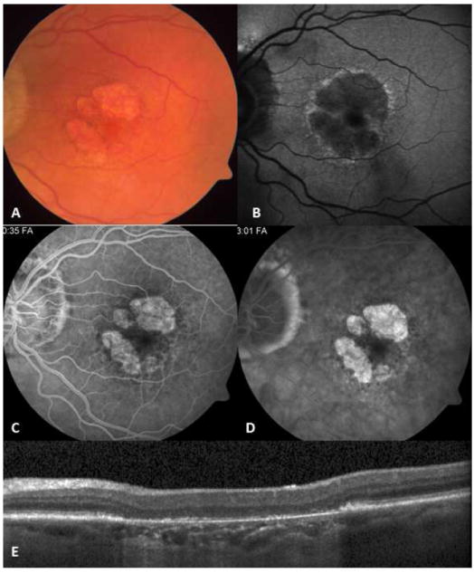
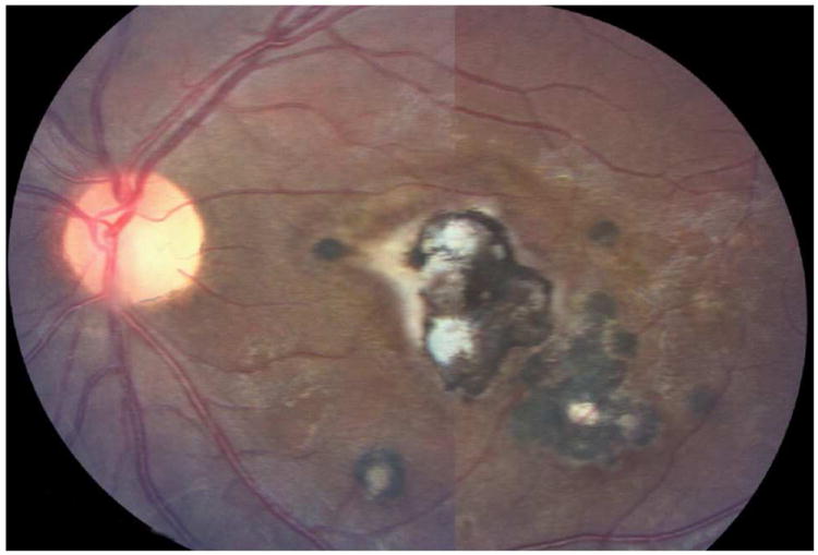
Serpiginous choroiditis (SC) is a posterior uveitis displaying a geographic pattern of choroiditis, extending from the juxtapapillary choroid and intermittently spreading centrifugally. The choroiditis involves the overlying retinal pigment epithelium, and the outer retina. This intraocular inflammation typically involves both eyes in otherwise healthy, middle-aged individuals with no familial or ethnic predilection. Pathogenesis is unclear; based on limited histopathologic studies, however, favorable response to immunosuppressive agents, and the absence of association with systemic or local infectious or noninfectious diseases, an organ-specific autoimmune inflammation seems likely to be the underlying process. Patients, particularly from tuberculosis-endemic regions, may present with fundus changes simulating SC, but show evidence of active tuberculosis and/or the presence of mycobacterial DNA in the aqueous humor. This has been referred to as serpiginous-like choroiditis, but we prefer the description multifocal serpiginoid choroiditis (MSC). We present the distinguishing features of SC and infectious multifocal serpiginoid choroiditis simulating SC. The distinction is crucial to avoid unnecessarily treating SC with antimicrobial agents. Advances in diagnostic and imaging modalities can help differentiate SC from MSC. Novel local and systemic treatment approaches improve the outcome and preserve vision in SC.


Figures

Figure 1
Figure 1
Jonathan Hutchinson (1828-1913), an English surgeon, ophthalmologist, dermatologist, and pathologist, described serpiginous choroiditis in a healthy individual, in a patient with tuberculosis lymphadenopathy, and in a patient with syphilis.
Figure 2
Figure 2
Macular serpiginous choroiditis in a 26-yr-old Caucasian man. Fundus photographs of the right (A) and left (C) eyes display geographic atrophy patches accompanied with pigment clumping. Mid-phase fluorescein angiogram of the right eye (B) shows geographic atrophy involving the retinal pigment epithelium and choroid with hyperpigmentation along the margins and at the fovea. Note border staining from choriocapillaris of adjacent choroid. Two patches of choroiditis reactivation are present at the temporal and superior margins of the macular lesion in the left eye (C - arrows). Early phase fluorescein angiogram (D) reveals blocked fluorescence corresponding to the active choroiditis patches in the left eye. (Images courtesy of Kumar Rao, MD, Saint Louis, MO)
Figure 3
Figure 3
67-yr-old Caucasian woman with bilateral active serpiginous choroiditis lesions (A, B). In the early phase, while retinal pigment epithelium is not damaged, lesions are underrepresented in fundus autofluorescence (C, D); however, fluorescein angiography (E-K) better revealed the extent of hypofluorescence corresponding to the choriocapillaris nonperfusion patches. Short arrows indicate hypofluorescent patches in the early phase (E). The initial hypo-perfused patches (short arrows in E) progressively stain and reveal more prominent hyperfluorescence in the borders in later frames. The right eye presents as macular serpiginous choroiditis with isolated patches not connected to the optic disc. Left eye demonstrates serpiginous choroiditis lesions superior to the optic disc and a separate macular lesion. Early hypofluorescent patches (J) of the active choroiditis stain progressively and become relatively hyperfluorescent in late phase angiogram (K-long arrow).
Figure 4
Figure 4
Fundus photograph (A) and early (B) and mid-phase (C) fluorescein angiograms from a 42-yr-old Caucasian woman with serpiginous choroiditis reveal choroiditis reactivation in the fovea. Vision has acutely declined to 5/200. A, while healed scars originating from the peripapillary area are characterized with RPE and choroidal atrophy, and pigment clumping, active choroiditis (short arrow) demonstrates gray-yellow discoloration of the retina. In fluorescein angiography, active choroiditis appears as a hypofluorescent patch with progressive marginal hyperfluorescence from the leaking vessels (long arrow). Healed lesions are surrounded by marginal staining (B,C), which is more prominent in late phases of the angiogram (arrowhead). (Images courtesy of Kumar Rao, MD, Saint Louis, MO)
Figure 5
Figure 5
76-yr-old Caucasian woman with serpiginous choroiditis. Fundus photographs show progressive expansion of the geographic choroiditis lesions. (This patient has been reported in part in reference .)
Figure 6
Figure 6
34-yr old Afghani woman with history of bilateral choroiditis that progressed rapidly after treatment with oral corticosteroids. PPD was positive and chest x-ray was negative. (A) Red free fundus photography of the right eye reveals geographic areas of choroidal atrophy, pigment clumping, and cystoid macular edema. (B) In fluorescein angiography, border staining delineates atrophic areas with some blockage from pigment proliferation. Early accumulation of dye in cystoid spaces is evident. (C, D) Mid-phase and late phase indocyanine green angiography reveals choriocapillaris loss in areas of healed lesions with preservation of large choroidal vessels. Choroidal circulation in non-involved areas is normal.
Figure 7
Figure 7
75-yr-old Caucasian woman with a documented history of contact with a patient with active pulmonary tuberculosis. She converted to PPD positive, and for this, she received anti-tuberculosis treatment. However, the number of drugs taken and the duration of treatment are unknown. Right and left eyes (A, B) fundus autofluorescent images; the autofluorescence is lost uniformly in atrophic areas. Note multifocal nature of the lesions that have coalesced in the center to form a larger patch. Satellite lesions are present. C and D correspond to mid-phase and late-phase fluorescein angiography and reveal a late staining of the exposed scleral bed and lesion borders. Optical coherence tomography scan (E) demonstrates loss of outer retina with pigment epithelial hypertrophy (arrow).
Figure 8
Figure 8
A 14-yr-old Caucasian woman with relentless placoid chorioretinitis (ampiginous choroiditis). (A) wide-field fundus autofluorescence image demonstrates multifocal inactive lesions distributed in the posterior pole and in the periphery of the left eye. Fundus photography (B) and late-phase fluorescein angiogram (C) reveal multifocal well-demarcated posterior pole lesions with late staining.
Figure 9
Figure 9
32-yr-old Asian Indian man with unilateral choroiditis that started in the macula and progressed rapidly following treatment with high dose oral corticosteroids. Tuberculosis work-up revealed positive QuantiFERON Gold test. Choroiditis subsided following institution of anti-tuberculosis treatment. Montage fundus photography (A) shows extensive healed lesions with pigmentary change in the posterior pole and in the periphery. Even though tuberculosis-associated multifocal serpiginoid choroiditis lesions are not connected to the peripapillary area at early presentation, advanced cases may present with extensive multifocal lesions involving the peripapillary area as well as the peripheral retina. There are two active patches of choroiditis in the supranasal periphery (arrow). Montage fluorescein angiogram (B) and wide-field autofluorescence image (C) reveal vividly the multifocal nature of the lesions.
Figure 10
Figure 10
A 24-yr-old Caucasian woman with idiopathic multifocal choroiditis. Left eye color photograph (A) and autofluorescent image (B) demonstrate multiple, round, atrophic lesions associated with pigment alterations in the posterior pole. The size and distribution of the lesions help in differentiation from larger and usually confluent patches seen in serpiginous choroiditis. Spectral domain-optical coherence tomography scan (C) reveals outer retinal destruction and pigment migration to the outer retina corresponding to healed chorioretinal lesions.
Figure 11
Figure 11
50-yr-old Caucasian man with presumed ocular histoplasmosis syndrome (POHS). Fundus photograph (A) and mid- and late angiograms (B, C) reveal multifocal punched-out atrophic lesions accompanied with peripapillary atrophy. Lesions are more prominent in fundus autofluorescence than in color fundus image (D). Spectral domain optical coherence tomography scan passing through the foveal lesion (E) shows a full-thickness destruction of the retina and choroid.
Figure 12
Figure 12
Nonexudative, age-related macular degeneration with geographic atrophy may resemble inactive serpiginous choroiditis lesion (A). In fundus autofluorescence imaging (B) a hyperautofluorescent halo surrounds the hypofluorescent lesion. Early window defect (C) due to atrophic retinal pigment epithelial (RPE) layer and late staining (D) of the atrophic bed with sharp margins are similar to angiographic features of inactive serpiginous choroiditis lesions. Optical coherence tomography scan (E) reveals atrophic RPE and increased backscattering from the choroid; however, outer retina retains normal reflectivity.
Figure 13
Figure 13
Toxoplasma chorioretinitis. Note a large geographic lesion within macula with satellite lesions.

References

    1. Abrams J, Schlaegel TF., Jr The tuberculin skin test in the diagnosis of tuberculous uveitis. Am J Ophthalmol. 1983;96(3):295–8. - PubMed
    1. Abrez H, Biswas J, Sudharshan S. Clinical profile, treatment, and visual outcome of serpiginous choroiditis. Ocul Immunol Inflamm. 2007;15(4):325–35. - PubMed
    1. Abu el-Asrar AM. Serpiginous (geographical) choroiditis. Int Ophthalmol Clin. 1995;35(2):87–91. - PubMed
    1. Adigüzel U, Sari A, Ozmen C, Oz O. Intravitreal triamcinolone acetonide treatment for serpiginous choroiditis. Ocul Immunol Inflamm. 2006;14(6):375–8. - PubMed
    1. Akpek EK, Baltatzis S, Yang J, Foster CS. Long-term immunosuppressive treatment of serpiginous choroiditis. Ocul Immunol Inflamm. 2001;9(3):147–67. - PubMed

Publication types

MeSH terms

LinkOut - more resources