Case Reports
. 2013 Apr 4;92(4):605-13.
doi: 10.1016/j.ajhg.2013.02.013. Epub 2013 Mar 28.

Perrault syndrome is caused by recessive mutations in CLPP, encoding a mitochondrial ATP-dependent chambered protease

Affiliations
Case Reports

Perrault syndrome is caused by recessive mutations in CLPP, encoding a mitochondrial ATP-dependent chambered protease

Emma M Jenkinson et al. Am J Hum Genet. .

Abstract

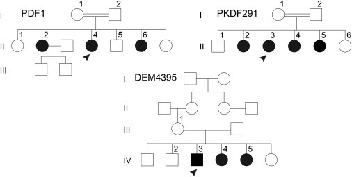
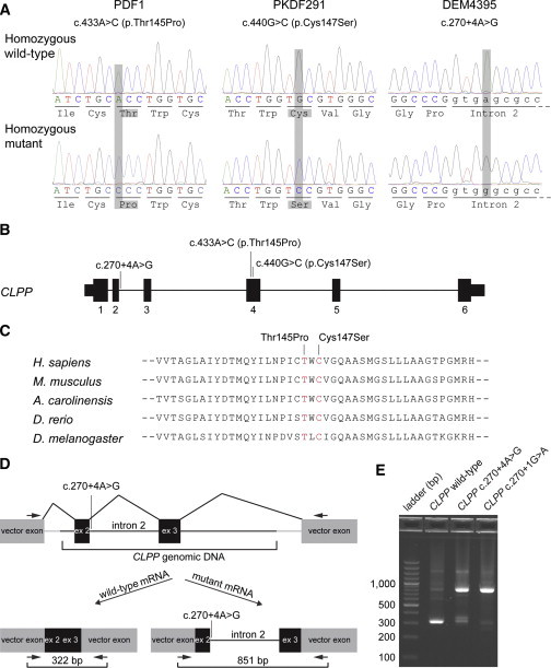
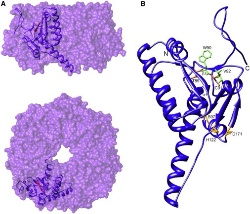
Perrault syndrome is a genetically and clinically heterogeneous autosomal-recessive condition characterized by sensorineural hearing loss and ovarian failure. By a combination of linkage analysis, homozygosity mapping, and exome sequencing in three families, we identified mutations in CLPP as the likely cause of this phenotype. In each family, affected individuals were homozygous for a different pathogenic CLPP allele: c.433A>C (p.Thr145Pro), c.440G>C (p.Cys147Ser), or an experimentally demonstrated splice-donor-site mutation, c.270+4A>G. CLPP, a component of a mitochondrial ATP-dependent proteolytic complex, is a highly conserved endopeptidase encoded by CLPP and forms an element of the evolutionarily ancient mitochondrial unfolded-protein response (UPR(mt)) stress signaling pathway. Crystal-structure modeling suggests that both substitutions would alter the structure of the CLPP barrel chamber that captures unfolded proteins and exposes them to proteolysis. Together with the previous identification of mutations in HARS2, encoding mitochondrial histidyl-tRNA synthetase, mutations in CLPP expose dysfunction of mitochondrial protein homeostasis as a cause of Perrault syndrome.


Figures

Figure 1
Figure 1
Pedigrees of the Three Families—PDF1, PKDF291, and DEM4395—Affected by Homozygous CLPP Mutations Numbers are assigned only to individuals whose DNA was available for this study. Arrowheads denote individuals whose genomic DNA was subjected to exome sequencing. Parents of the six siblings in family PKDF291 have the same great-great grandparents. A double horizontal line denotes a consanguineous union. Family PDF1 was ascertained because of profound hearing loss in three sisters with subsequent POF. Families PKDF291 and DEM4395 first came to attention because of profound hearing loss in the affected family members. In family PKDF291, POF was revealed by subsequent evaluation of the affected sisters. In family DEM4395, no hormonal evaluation was possible.
Figure 2
Figure 2
Identification of CLPP Mutations in Families PDF1, PKDF291, and DEM4395 (A) Chromatograms obtained from a wild-type control and homozygous affected individuals from three families. Mutations are highlighted in gray. (B) Gene structure of CLPP and the location of the three mutations identified in this study. Thin bars represent 5′ and 3′ UTRs, and thick bars represent exons. The horizontal lines joining exons represent intronic sequence. (C) A ClustalW alignment of CLPP orthologs in five animal species from human to Drosophila shows conservation of residues 145 and 147, which correspond to the substitutions identified in families PDF1 and PKDF291, respectively. (D) Schematic presentation of the exon-splicing-assay vector used in COS-7 cells for the evaluation of the predicted splice-site mutation. CLPP exon 2, either with a wild-type donor site or a mutant donor splice site (c.270+4A>G or control c.270+1G>A), was cloned into the pSPL3 expression vector (gray). The CLPP sequence also included intron 2, exon 3, and 660 bp of flanking intronic sequences (black). Horizontal arrows indicate locations of vector-specific primers used for PCR amplification of cDNA containing the CLPP sequence. (E) Splicing-assay products were separated by size on a 2% agarose gel for the wild-type allele, the DEM4395 c.270+4A>G mutant allele, and the c.270+1G>A control mutation. The 851 bp band is from CLPP transcripts that include intron 2. Sequencing of the two ∼300–350 bp gel-purified bands demonstrates that the c.270+4A>G mutant allele results in some wild-type splicing between CLPP exons 2 and 3. Additional splicing assay data in Table S2 support this conclusion.
Figure 3
Figure 3
Location of Substitutions within the Crystal Structure of Human CLPP (A and B) Surface representations show the side (A) and top (B) views of a single heptameric ring of CLPP subunits (Protein Data Bank 1tg6). The ribbon representation shows a single monomer within the ring in each case. The β strand affected by the two substitutions is highlighted in red. (B) A ribbon representation shows the position of substitutions at the base of the CLPP hydrophobic pocket. Substitutions are shown in red, and adjacent hydrophobic residues known to be important in CLPX binding are shown in green. The catalytic triad is highlighted in orange. The single-letter codes for amino acid residues and numbers shown here are after cleavage of the mitochondrial targeting sequence (MTS), which removes the N-terminal 56 residues. Hence, the residue labeled T89 is equivalent to T145 in the unprocessed polypeptide. This is for consistency with studies relating to the structure of CLPP. The poorly resolved N-terminal region (residues 1–17) was omitted for clarity.
Figure 4
Figure 4
Immunolocalization of CLPP in Human Fetal Ovary, Adult Mouse Ovary, and P3 Mouse Organ of Corti (A) Immunodetection (brown staining) of CLPP in germ cells of human fetal ovary (approximately 18 weeks of gestation; 20 mm foot length). Staining was particularly evident around nuclei of germ cells, consistent with the localization of mitochondria. Staining was not evident in the stromal layers. Human fetal tissue was collected with ethical approval under the Codes of Practice of the UK Human Tissue Authority and staged by foot length. For bright-field studies, endogenous peroxidase was quenched by incubation with 30% hydrogen peroxide and antigen retrieval was undertaken by heating at 95°C for 5 minutes in sodium citrate pH 6. Rabbit CLPP antibody (Sigma-Aldrich HPA010649) was used as the primary antibody, and unconjugated goat anti-rabbit IgG (Vector Labs AI-1000) was used as the secondary antibody. A system of streptavidin, horseradish peroxidase, and diaminobenzidine (Vector Labs) was used for generating a brown signal. Scale bars represent 50 μm. (B) Immunoblot analysis for the validation of the rabbit monoclonal CLPP antibody (AbCam ab124822). When used at a 1:2,000 dilution in ECL Prime blocking reagent (GE RPN418V), this rabbit monoclonal antibody recognizes a predicted 26 kDa CLPP protein in 4 μg of a postnatal day (P) 25 mouse ovary lysate, 4 μg of a P3 mouse cochlea lysate, and 0.5 μg of COS-7 cell lysate. COS-7 cells were also transfected with pAcGFP-N2-CLPP and expressed GFP-tagged CLPP proteins of 57 kDa and 51 kDa. The latter protein presumably lacks the predicted 6 kDa MTS. Endogenous 26 kDa CLPP was also detected in these COS-7 cells. (C and D) Immunohistochemistry of an adult mouse ovary reveals CLPP expression in granulosa cells (G) and oocytes (O). CLPP staining is absent in the zona pellucida (ZP) and theca (T). Fixed-frozen sections (Zyagen MF-406) were labeled with a rabbit monoclonal CLPP antibody as described below and counterstained with a 1:100 dilution of phalloidin-Atto 390 (Sigma 50556). Scale bars represent 30 μm. (E) An optical section of a P3 mouse organ of Corti at the level of the cuticular plate reveals that CLPP (green) is more abundant in Deiter’s cells (DCs), inner pillar cells (PCs), and Hensen’s cells (HCs) than in inner sulcus cells (ISCs), inner hair cells (IHCs), and outer hair cells (OHCs). CLPP colocalized with the mitochondrial protein cytochrome c (red). After dissection from the temporal bone, cochleae were fixed in 4% paraformaldehyde. Tissues were finely dissected, permeabilized in PBS containing 0.5% Triton X-100, and incubated overnight in 1:100 dilutions of rabbit CLPP antibody and mouse cytochrome c antibody (BD Biosciences 556433) in 5% goat serum and 2% BSA blocking solution. Primary antibodies were detected with 1:400 dilutions of Alexa Fluor 488 donkey anti-rabbit IgG (Invitrogen 21206) and Alexa Fluor 568 goat anti-mouse IgG (Invitrogen 11004) in blocking solution. Scale bars represent 10 μm.

References

    1. Guest S.S., Evans C.D., Winter R.M. The Online London Dysmorphology Database. Genet. Med. 1999;1:207–212. - PubMed
    1. Lenz D.R., Avraham K.B. Hereditary hearing loss: from human mutation to mechanism. Hear. Res. 2011;281:3–10. - PubMed
    1. Perrault M., Klotz B., Housset E. Deux cas de syndrome de Turner avec surdi-mutite dans une meme fratrie. Bull. Mem. Soc. Med. Hop. Paris. 1951;16:79–84. - PubMed
    1. Fiumara A., Sorge G., Toscano A., Parano E., Pavone L., Opitz J.M. Perrault syndrome: evidence for progressive nervous system involvement. Am. J. Med. Genet. A. 2004;128A:246–249. - PubMed
    1. Jenkinson E.M., Clayton-Smith J., Mehta S., Bennett C., Reardon W., Green A., Pearce S.H., De Michele G., Conway G.S., Cilliers D. Perrault syndrome: further evidence for genetic heterogeneity. J. Neurol. 2012;259:974–976. - PubMed

Publication types

MeSH terms

Supplementary concepts

LinkOut - more resources