. 2013 Apr 8;23(7):560-8.
doi: 10.1016/j.cub.2013.02.047. Epub 2013 Mar 28.

Electrophoresis of cellular membrane components creates the directional cue guiding keratocyte galvanotaxis

Affiliations

Electrophoresis of cellular membrane components creates the directional cue guiding keratocyte galvanotaxis

Greg M Allen et al. Curr Biol. .

Abstract

Background: Motile cells exposed to an external direct current electric field will reorient and migrate along the direction of the electric potential in a process known as galvanotaxis. The underlying physical mechanism that allows a cell to sense an electric field is unknown, although several plausible hypotheses have been proposed. In this work we evaluate the validity of each of these mechanisms.

Results: We find that the directional motile response of fish epidermal cells to the cathode in an electric field does not require extracellular sodium or potassium, is insensitive to membrane potential, and is also insensitive to perturbation of calcium, sodium, hydrogen, or chloride ion transport across the plasma membrane. Cells migrate in the direction of applied forces from laminar fluid flow, but reversal of electro-osmotic flow did not affect the galvanotactic response. Galvanotaxis fails when extracellular pH is below 6, suggesting that the effective charge of membrane components might be a crucial factor. Slowing the migration of membrane components with an increase in aqueous viscosity slows the kinetics of the galvanotactic response. In addition, inhibition of PI3K reverses the cell's response to the anode, suggesting the existence of multiple signaling pathways downstream of the galvanotactic signal.

Conclusions: Our results are most consistent with the hypothesis that electrophoretic redistribution of membrane components of the motile cell is the primary physical mechanism for motile cells to sense an electric field. This chemical polarization of the cellular membrane is then transduced by intracellular signaling pathways canonical to chemotaxis to dictate the cell's direction of travel.


Figures

Figure 1
Figure 1. Models for directional sensing of a keratocyte in an electric field
Visual description of the possible models for a galvanotactic response of a motile cell. (A) An electric field will polarize the cell changing electro-motive forces and opening/closing voltage gated ion channels. (B) Electro-osmotic flow, νeo, at the charged migration surface will apply external force on the cell, which could for instance displace adhesions laterally. (C) Electro-osmotic forces created by the relatively immobile charged ions in the cell membrane attracting a mobile double layer at the cell surface will combine with electro-static forces on charged macromolecules and membrane components to produce mechanical work. As depicted, this could asymmetrically activate a force sensor creating a local signal that could be used to define the front and the back of the cell. (D) Local electro-osmotic and electrostatic forces at the cell membrane will also electrophorese membrane components. Negatively charged components will move to the anode, positively charged components will migrate to the cathode. Electro-osmotic forces at the membrane will also act to push proteins to one side of the cell or the other depending on the net surface charge of the cell.
Figure 2
Figure 2. Keratocytes migrate to the cathode in an electric field
(a) Rose plots of the distribution of angles traveled in populations of 137 control cells (p=0.56) and (b) 110 cells in an electric field of 10 V/cm (p = 5.9 × 10⁻⁹), with the cathode oriented toward the right. The p value is calculated from a Kolmogorov-Smirnov test for a uniform distribution of angles traveled. (c) The strength of the directional response is calculated for populations of cells as the mean ± standard error of the cos(θ), where θ represents the direction that a cell travels relative to the electric field lines, as depicted graphically. A cos(θ) of 1 indicates a complete directional response towards the cathode (purple line), a cos(θ) of 0 indicates no response (cyan line), and a cos(θ) of −1 indicates a reversed response. Green points and fit line represent the dose response to the applied potential of cells in normal media. For a given applied potential, there was a decreased strength of response when media conductivity was decreased by mixing L-15 media 1:4 (red) or 1:10 (blue) with water. (d) Re-plotting the same data as in panel c in terms of current flow shows that for all salt concentrations the directional response is proportional to the ionic current. Given the constant flow cell geometry used in these analysis, current density, J, will depend on the measured current, I, and cross-sectional area of the flow cell, A, where J=IA=I2×107m2
Figure 3
Figure 3. Ionic flux does not drive galvanotaxis
Direction of travel of cells as quantified by the cos(θ) (blue) and mean cell speed (red) for populations of cells under specified perturbations, with n indicating number of cells analyzed and error bars indicating the standard error of the mean. All perturbations were performed under an electric field of 5 V/cm (1.5 mA) except for the no electric-field control cells. Perturbations include Na+ free salt solution (Na+ ions replaced with K+ ions) [similar results were seen with Cs+ supplementation], K+ free media (Na+ supplemented), Ca++ ionophore A-21387, intracellular Ca++ chelator BAPTA-AM, L-type voltage gated Ca++ channel blocker verapamil, epithelial sodium channel (ENaC) and Na+/H+ exchanger (NHE-1) inhibitor amiloride, volume regulated anion channel (VRAC) inhibitors DCPIB and quinine, ATP and ADP (which can act as chloride channel inhibitors), vacuolar-type H+-ATPase inhibitor bafilomycin, and proton ionophore dinitrophenol.
Figure 4
Figure 4. Keratocytes migrate in the direction of applied force, but bulk electro-osmotic flow does not drive galvanotaxis
(c) Measured flow profile seen when a 5 V/cm electric field exists across the flow cell as measured by fluorescent tracer particles. Negligible particle motion is noted when the electric field is off. Re-circulation flow in the center of the flow cell was noted due to the static head of pressure at the ends of the flow cell. Control conditions (left, red) produced flow toward the cathode. Coating surfaces with 20 μg/mL poly-l-lysine (right, blue) reversed the direction of electro-osmotic flow to the anode. Cartoon keratocyte is not drawn to scale. (d) Rose plot of directions of travel of keratocytes under an electric field of 1 V/cm (0.33 mA) with the cathode to the right under control conditions (left) and with the substrate patterned with 20 μg/mL poly-l-lysine (right). Cells exhibited a robust galvanotactic response to the cathode under both conditions.
Figure 5
Figure 5. Keratocyte galvanotaxis is sensitive to protonation
(a) Measured cos(θ) (green) and cell speeds (magenta) ± standard error for cells in media of variable pH when exposed to an electric field of 5 V/cm. Cells retained robust motility across this pH range. All measurements were done without the presence of serum, except pH of 5.8, 6.2 and 7.4. The presence of serum did not effect directionality at an acidic pH. (b) The mean ± standard error of the electrophoretic mobility of red blood cells and keratocytes in suspension and in an electric field of 1.5 V/cm. Red blood cells phoresed towards the anode as would be expected from a negatively charged cell, keratocytes phoresed towards the cathode as would be expected from a positively charged cell. Note the electrophoretic mobility speed is an order of magnitude larger than typical speeds of cell migration.
Figure 6
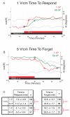
Figure 6. Time to Respond and Time to Forget are dependent on aqueous viscosity
The calculated mean(cos(θ)) is shown at every time point for all observed cells. Dashed black line represents control cells at steady state in the electric field, dashed blue line represents control cells at steady state without an electric field, red line indicates cells in 1 cP media and green line represents cells in 50 cP media + methyl-cellulose. (a) Graphical depiction of the time of cells to respond to a 1 V/cm (0.3 mA) electric field, where the electric field is turned on at the 15th minute for cells in 1 and 50 cP media. We see cells in the lower viscosity media reach steady state behavior (red circle) faster than cells in higher viscosity media (green circle). (b) Graphical depiction of the time of cells to forget a 5 V/cm (1.5 mA) electric field, where the electric field is turned off at the 30th minute for cells in 1 and 50 cP. Again we see cells in the lower viscosity media reach the new steady state behavior (red circle) faster than cells in higher viscosity media (green circle). (c) The mean ± standard error of the time to respond and time to forget were quantified from the time it takes each cell to reach steady state in minutes. The p-value of unpaired Student’s two-sample t-tests between normal and elevated viscosity are marked in red for each.
Figure 7
Figure 7. Keratocyte galvanotaxis is sensitive to PI3K activity
Rose plots of the distribution of angles traveled in populations of cells exposed to electric field of 1 and 5 V/cm (cathode oriented to the right) with and without the presence of PI3K inhibitor LY-294002. The fraction of cells traveling to the either the quartile of angles representing the cathode or anode are represented in red. Inhibition of PI3K causes some cells at 1 V/cm and a majority of cells at 5 V/cm to to reverse direction to the anode.

References

    1. Verworn M. Die polare Erregung der Protisten durch den galvanischen strom. (“The polar excitation of protists by galvanic current”) Pflugers Arch Ges Physiol. 1889;45:1–36.
    1. Hotary KB, Robinson KR. Endogenous electrical currents and voltage gradients in Xenopus embryos and the consequences of their disruption. Dev Biol. 1994;166(2):789–800. doi:10.1006/dbio.1994.1357. - PubMed
    1. McCaig CD, Song B, Rajnicek AM. Electrical dimensions in cell science. J Cell Sci. 2009;122(Pt 23):4267–76. doi:10.1242/jcs.023564. - PubMed
    1. Zhao M. Electrical fields in wound healing-An overriding signal that directs cell migration. Semin Cell Dev Biol. 2009;20(6):674–82. doi:10.1016/j.semcdb.2008.12.009. - PubMed
    1. McCaig CD, Rajnicek AM, Song B, Zhao M. Controlling cell behavior electrically: current views and future potential. Physiol Rev. 2005;85(3):943–78. doi:10.1152/physrev.00020.2004. - PubMed

Publication types

Substances