. 2014 Mar 13;33(11):1438-47.
doi: 10.1038/onc.2013.78. Epub 2013 Apr 1.

Pin1 modulates ERα levels in breast cancer through inhibition of phosphorylation-dependent ubiquitination and degradation

Affiliations

Pin1 modulates ERα levels in breast cancer through inhibition of phosphorylation-dependent ubiquitination and degradation

P Rajbhandari et al. Oncogene. .

Abstract

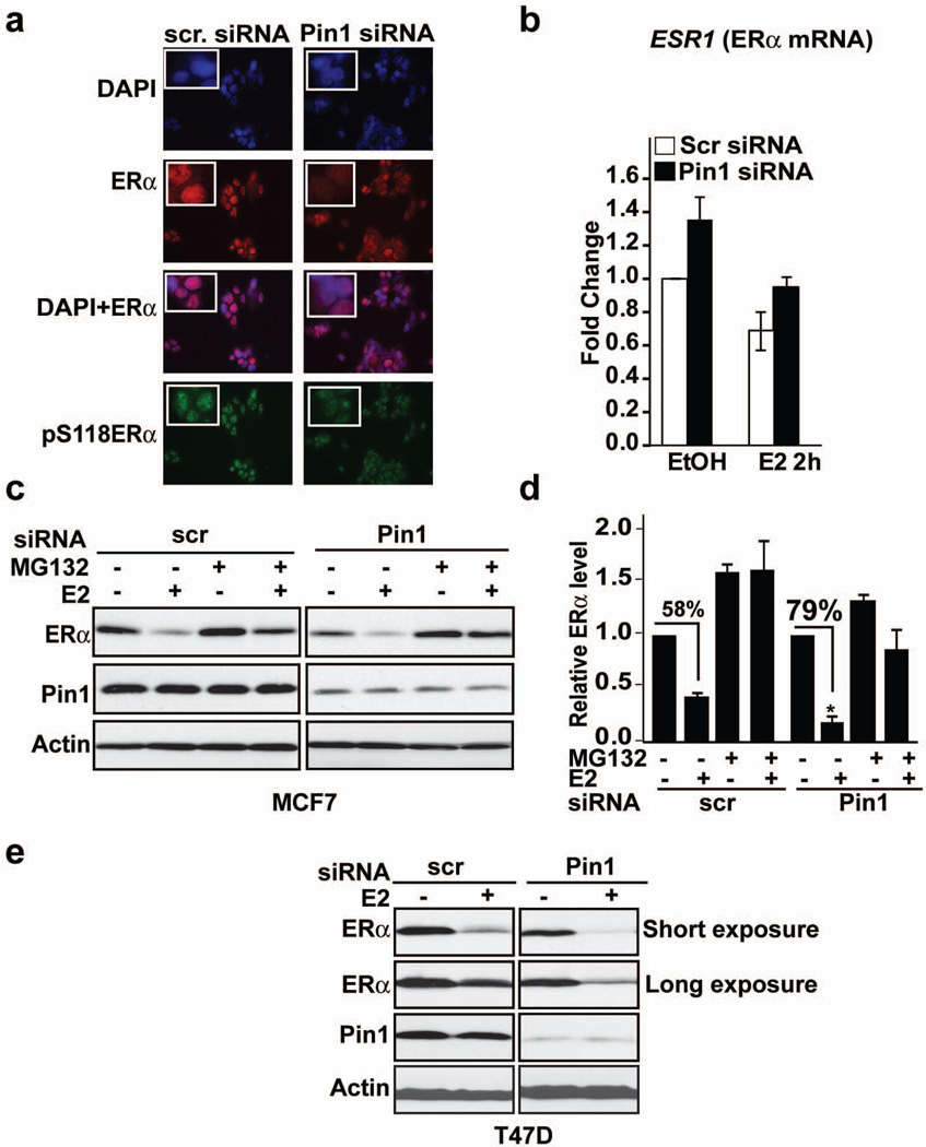
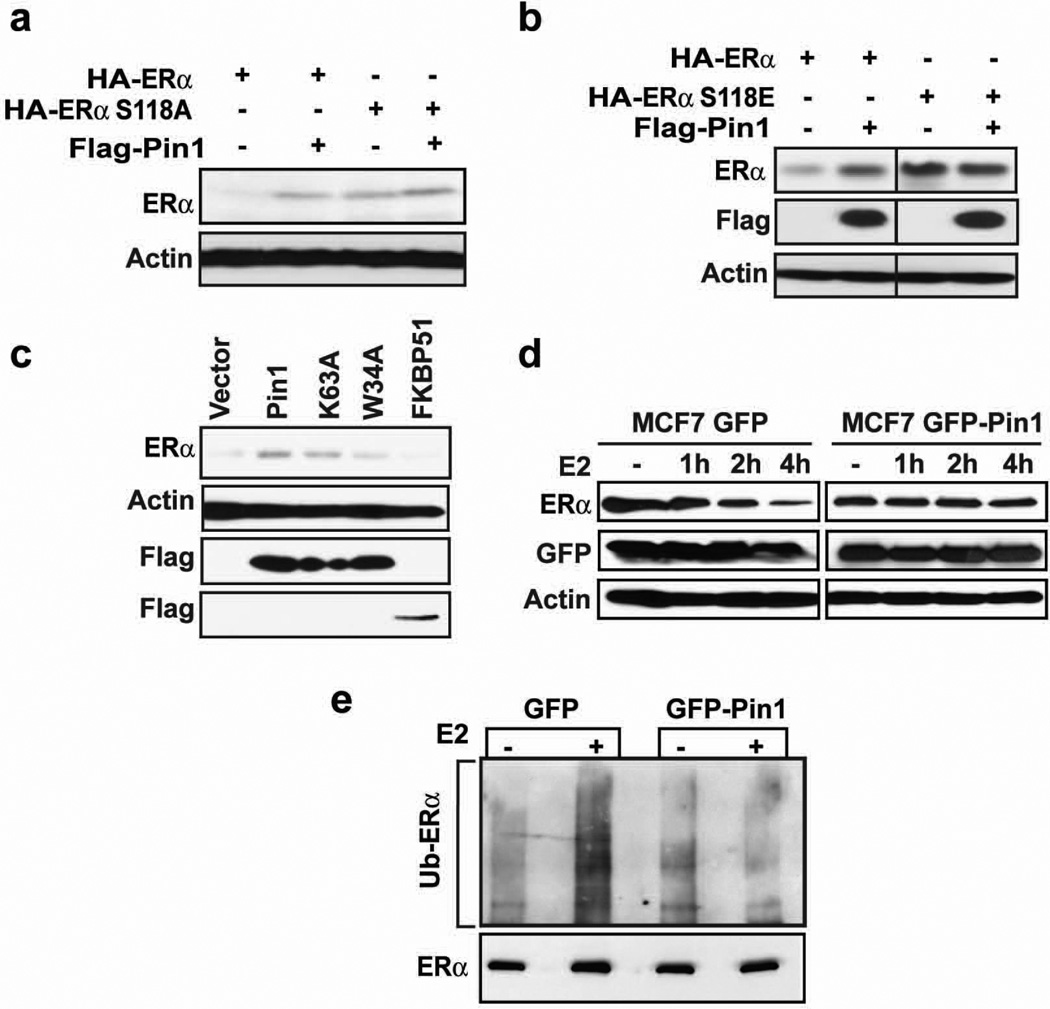
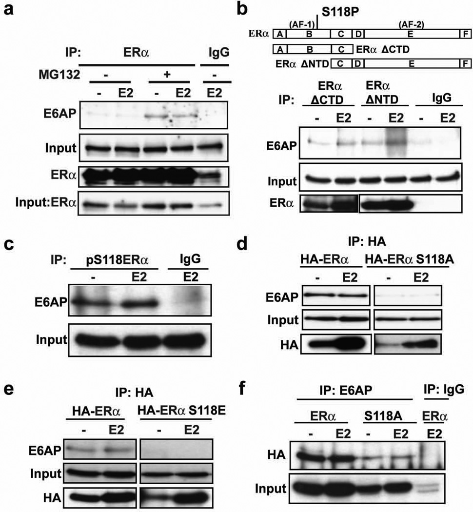
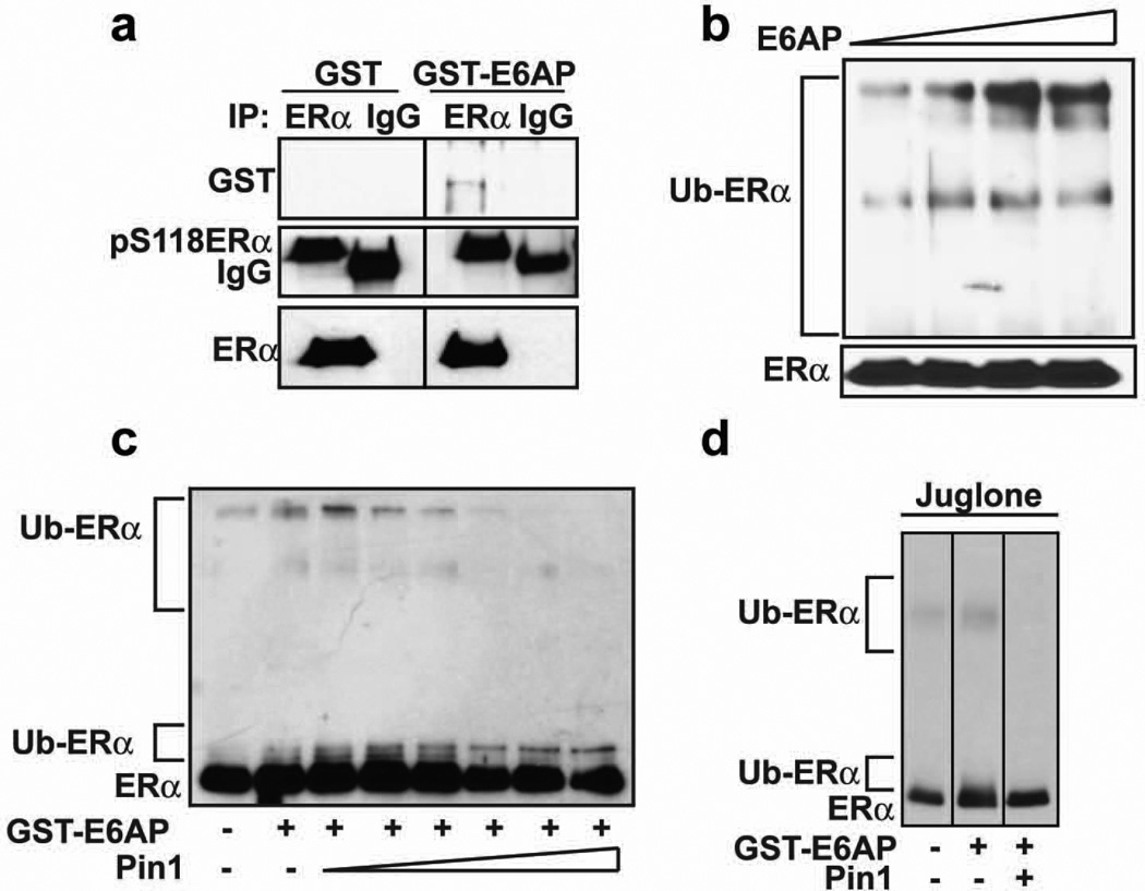
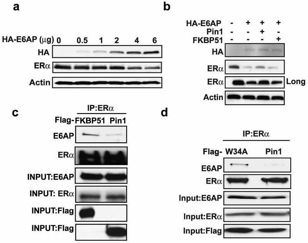
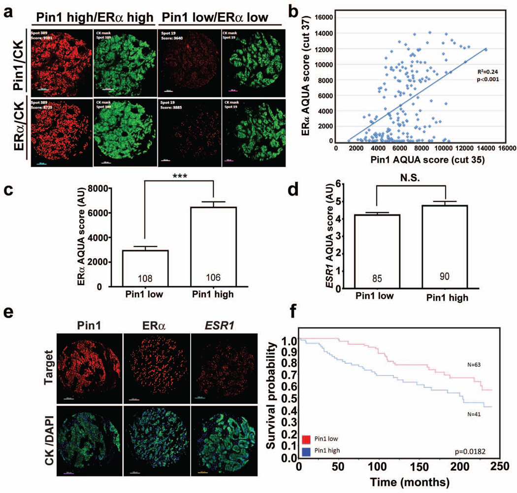
Estrogen receptor-alpha (ERα) is an important biomarker used to classify and direct therapy decisions in breast cancer (BC). Both ERα protein and its transcript, ESR1, are used to predict response to tamoxifen therapy, yet certain tumors have discordant levels of ERα protein and ESR1, which is currently unexplained. Cellular ERα protein levels can be controlled post-translationally by the ubiquitin-proteasome pathway through a mechanism that depends on phosphorylation at residue S118. Phospho-S118 (pS118-ERα) is a substrate for the peptidyl prolyl isomerase, Pin1, which mediates cis-trans isomerization of the pS118-P119 bond to enhance ERα transcriptional function. Here, we demonstrate that Pin1 can increase ERα protein without affecting ESR1 transcript levels by inhibiting proteasome-dependent receptor degradation. Pin1 disrupts ERα ubiquitination by interfering with receptor interactions with the E3 ligase, E6AP, which also is shown to bind pS118-ERα. Quantitative in situ assessments of ERα protein, ESR1, and Pin1 in human tumors from a retrospective cohort show that Pin1 levels correlate with ERα protein but not to ESR1 levels. These data show that ERα protein is post-translationally regulated by Pin1 in a proportion of breast carcinomas. As Pin1 impacts both ERα protein levels and transactivation function, these data implicate Pin1 as a potential surrogate marker for predicting outcome of ERα-positive BC.


Conflict of interest statement

CONFLICT OF INTEREST

The authors declare no conflict of interest.

Figures

Figure 1
Figure 1. Loss of Pin1 accelerates proteasome-mediated degradation of ERα
a) Immunofluorescence microscopy of MCF-7 cells transfected with Pin1 siRNA or control scrambled (scr) siRNA and treated with E2 for 2h. Fixed cells were incubated with an anti-ERα or anti-pS118ERα antibody and stained with DAPI for nuclear staining. b) MCF-7 cells were transfected with Pin1 siRNA or control scr siRNA. The cells were treated with EtOH or E2 for 2 h, and qRT-PCR was used to analyze the expression of ERα mRNA, ESR1. Data shown are relative to those of the EtOH- treated control (left-most bar), and data are represented as means +/- SEM for three independent experiments. c and d) MCF-7 cells were transfected with Pin1 siRNA or control scr siRNA, and 72 h after transfection, cells were pretreated with and without 10 µM MG132, a proteasome inhibitor, for 30 min followed by EtOH (−) or 10 nM E2 treatment for 2 h. Levels of ERα, Pin1, and actin (loading control) were assessed by Western blot analysis and (d) bands were quantified by densitometry and represented as a graph normalized to EtOH-treated samples. Data are represented as means +/− SEM for three independent experiments. Asterisks indicate a statistically significant difference between scr siRNA and Pin1 siRNA E2-treated cells (*, p < 0.05), using Student’s t test, and % denotes changes between EtOH and E2-treated samples. e) T47D cells were transfected as in (c) and treated with EtOH (−) or 10 nM E2 (+) for 2h. Levels of ERα, Pin1, and actin were assessed by Western blot analysis.
Figure 2
Figure 2. Pin1 stabilizes ERα in an S118-dependent manner and blocks its ubiquitination
a and b) MEF Pin1−/− were co-transfected with 0.1 µg Flag-Pin1 and 0.3 µg of HA-ERα or HA-ERα S118A (a) or HA-ERα S118E (b) for 24 h and treated with 10 nM E2 for another 24 h. Western blot analysis was performed to assess the level of ERα by using anti-HA antibody and Pin1 by anti-Flag antibody. The actin band represents the loading control. c) MEF Pin1−/− cells were co-transfected with HA-ERα and Flag vector, Flag-Pin1, Flag-Pin1 K63A, Flag-Pin1 W34A, or Flag-FKBP51. 24 h post-transfection, cells were treated with 10 nM E2 for 24 h and Western blot was performed for ERα, Flag, and actin. d) MCF-7 cells stably expressing GFP or GFP-Pin1 were treated with and without 10 nM E2 for the indicated length of time and Western blot was performed for ERα, GFP, and actin. e) MCF-7 cells overexpressing GFP or GFP Pin1 were treated with 10 µM MG132 for 30 min followed by 4 h treatment with 10 nM E2 (+) or EtOH (-). ERα was immunoprecipitated using anti-ERα antibody and the level of ubiquitination was evaluated by Western blot using anti-ubiquitin antibody (Ub).
Figure 3
Figure 3. The E3 ligase, E6AP, binds ERα in a S118-dependent manner
a) MCF-7 cells were pre-treated with or without 10 µM MG132 for 30 mins and then treated with and without 10 nM E2 and 10 µM MG132 for 4 h. Cells were harvested and ERα was immunoprecipitated by ERα antibody or normal rabbit IgG, and Western blot was performed for E6AP and ERα. Input lanes represent E6AP and ERα in cell extracts before immunoprecipitation. b) Schematic representation of regions corresponding to full length ERα, ERα ΔCTD, and ERα ΔNTD. Also shown is the location of AF1, AF2 and S118P sites (Upper panel). 293T cells were transfected with plasmid HE15 expressing ERα ΔCTD or HE19 expressing ERα ΔNTD. 24 h post-transfection, cells were treated as in (a) and ERα ΔCTD was immunoprecipitated with N-terminus specific ERα antibody (H184, Santa Cruz Biotechnology) and ERα ΔNTD with C-terminus specific ERα antibody (HC20, Santa Cruz Biotechnology), or rabbit IgG, and Western blot was performed with respective ERα antibodies and E6AP. c) MCF-7 cells were treated as in (a) and immunoprecipitated with pS118-ERα antibody or mouse IgG and Western blot was performed for E6AP. d and e) 293 cells stably expressing HA-ERα, HA-ERα S118A (d) or HA-ERα S118E (e) were treated as in (a) and ERα was immunoprecipitated with HA antibody and Western blot was performed for E6AP and HA. f) 293 cells stably expressing HA-ERα or HA-ERα S118A were treated as in (d) and E6AP was immunoprecipitated with E6AP antibody or rabbit IgG and Western blot was performed for HA.
Figure 4
Figure 4. Pin1 prevents E6AP-mediated in vitro ubiquitination of ERα
a) Purified recombinant ERα and GST or GST-E6AP were allowed to form complexes for 1 h, and then ERα was immunoprecipitated using ERα antibody or mouse IgG antibody and Western blot performed for GST, pS118ERα, and ERα. b) ERα was in vitro ubiquitinated as described in the experimental procedures with Ube1, UbcH5a, ATP, ubiquitin, and GST-E6AP (0–0.6 µg) at 30°C for 1.5 h, and Western blot was performed for ERα. c) ERα was in vitro ubiquitinated as in (b) with and without 0.4 µg GST-E6AP in the presence and absence of Pin1 (0–1 µg), and Western blot was performed for ERα. d) ERα was ubiquitinated as in (c) with and without GST-E6AP and Pin1 in the presence and absence of juglone (7.5 µM), and Western blot was performed for ERα.
Figure 5
Figure 5. Pin1 blocks E6AP and ERα interaction
a) 293T cells were transfected with and without HA-E6AP (0–6 µg) and ERα for 24 h and Western blot was performed for HA, ERα, or actin. b) 293T cells were co-transfected with and without HA-E6AP, ERα and Flag vector, Flag-Pin1, Flag-FKBP51 for 24 h, and Western blot was performed for HA, ERα, and actin. The long exposure represents an increase in the length of time the blot was exposed to X-ray film c) MCF-7 cells were transfected with Flag-FKBP51 or Flag-Pin1, and 24 h post-transfection, cells were pretreated with 10 µM MG132 for 30 mins and with 10 nM E2 and 10 µM MG132 for 4h. Cells were then lysed and extracts were immunoprecipitated for ERα and Western blot was performed for E6AP, ERα, and Flag. Input lanes show proteins in cell extracts before immunoprecipitation. d) MCF-7 cells were transfected with Flag-Pin1 or Flag-Pin1 W34A for 24 h and immunoprecipitation and Western blot was performed as in (c).
Figure 6
Figure 6. Pin1 protein expression in human breast carcinomas and relationship with ERα and ESR1 levels
a) Fluorescence microphotograph showing representative cases with high Pin1 and ERα scores and with low Pin1 and ERα on a Yale Pathology cohort (YTMA-201). On the right upper and lower panels (green channel) are the corresponding pancytokeratin (CK) masks used for AQUA analysis. b) Relationship between Pin1 and ERα in samples from YTMA201. Pin1 and ERα were measured using AQUA in serial sections as indicated by the cut number in parentheses. R2 indicates linear regression coefficient between scores. Pin1 and ERα protein showed significant correlation with p<0.001. c and d) Average ERα (c) and ESR1 mRNA levels (d) in BC samples from YTMA201 showing low or high Pin1 levels. Pin1 low cases include those scores below the median; and Pin1 high include those above the median AQUA score. Number of cases in each group is indicated within each bar. ***=p<0.001 (U-test and Mann-Whitney); NS= not significant. e) Representative fluorescence microphotograph showing Pin1, ERα, and ESR1 expression in human BC samples on YTMA201. The lower panels (green and blue channels) show the corresponding pancytokeratin (CK) and nuclear DAPI stainings. f) Pin1 protein expression is associated with survival in human breast cancer. Kaplan-Meier curves showing 20-year overall survival probability of Pin1 high (blue line) and Pin1 low (red line) ERα positive breast cancer patients from the retrospective Yale TMA 201cohort.

References

    1. Dowsett M, Cuzick J, Ingle J, et al. Meta-analysis of breast cancer outcomes in adjuvant trials of aromatase inhibitors versus tamoxifen. J Clin Oncol. 2010;28:509–518. - PubMed
    1. Early Breast Cancer Trialists' Collaborative Group EBCTCG. Effects of chemotherapy and hormonal therapy for early breast cancer on recurrence and 15-year survival: an overview of the randomised trials. The Lancet. 2005;365:1687–1717. - PubMed
    1. Harrell JC, Dye WW, Allred DC, et al. Estrogen receptor positive breast cancer metastasis: altered hormonal sensitivity and tumor aggressiveness in lymphatic vessels and lymph nodes. Cancer Res. 2006;66:9308–9315. - PubMed
    1. Harrell JC, Dye WW, Harvell DM, et al. Estrogen insensitivity in a model of estrogen r eceptor positive breast cancer lymph node metastasis. Cancer Res. 2007;67:10582–10591. - PubMed
    1. Khan SA, Rogers MA, Khurana KK, Meguid MM, Numann PJ. Estrogen receptor expression in benign breast epithelium and breast cancer risk. J Natl Cancer Inst. 1998;90:37–42. - PubMed

Publication types

Substances