Review
. 2013 Jul-Aug;164(6):620-39.
doi: 10.1016/j.resmic.2013.03.012. Epub 2013 Mar 28.

The expanding bacterial type IV secretion lexicon

Affiliations
Review

The expanding bacterial type IV secretion lexicon

Minny Bhatty et al. Res Microbiol. 2013 Jul-Aug.

Abstract

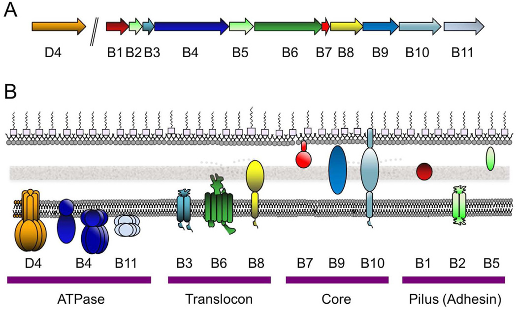
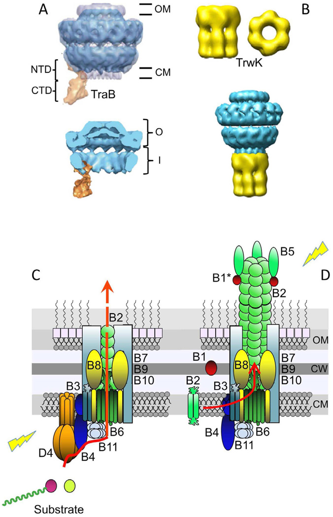
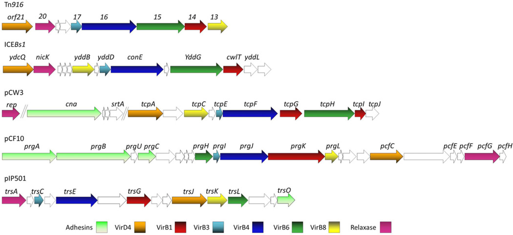
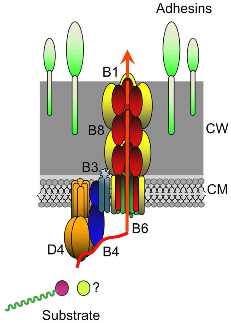
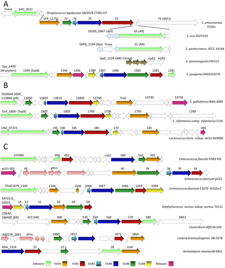
The bacterial type IV secretion systems (T4SSs) comprise a biologically diverse group of translocation systems functioning to deliver DNA or protein substrates from donor to target cells generally by a mechanism dependent on establishment of direct cell-to-cell contact. Members of one T4SS subfamily, the conjugation systems, mediate the widespread and rapid dissemination of antibiotic resistance and virulence traits among bacterial pathogens. Members of a second subfamily, the effector translocators, are used by often medically-important pathogens to deliver effector proteins to eukaryotic target cells during the course of infection. Here we summarize our current understanding of the structural and functional diversity of T4SSs and of the evolutionary processes shaping this diversity. We compare mechanistic and architectural features of T4SSs from Gram-negative and -positive species. Finally, we introduce the concept of the 'minimized' T4SSs; these are systems composed of a conserved set of 5-6 subunits that are distributed among many Gram-positive and some Gram-negative species.

Keywords: ATPase; Conjugation; Pathogenesis; Pilus; Translocation; Type IV secretion.


Figures

Fig. 1
Fig. 1
The A. tumefaciens VirB/VirD4 T4SS gene organization and subunit subcellular locations. (A) virD4 is co-transcribed with the virD1 relaxase and virD2 accessory factor genes from the virD promoter and the eleven virB genes are transcribed from the virB promoter. (B) VirD4 and VirB proteins are associated with the cytoplasmic membrane (CM), cell-wall (CW) containing periplasm, or outer membrane (OM) as shown. General membrane topologies are denoted as are the oligomeric structures of the VirD4 (hexamer), VirB4 (monomer or hexamer, see text), VirB11 (hexamer) ATPases. VirB7 is an outer-membrane-associated lipoprotein and VirB10 spans both membrane with N- and C-proximal α-helical domains. The VirD4 and VirB subunits are clustered into four functional groups.
Fig. 2
Fig. 2
Gram-negative T4SS subassemblies and architectural models. (A) Upper: Structure of the pKM101-encoded core complex with the VirB4-like TraB homolog bound to the side. The core is shown in blue and TraB is in orange. The O and I layers are indicated as O and I, respectively. TraBN-terminal domain (NTD) and C-terminal domain (CTD) are shown. The negative stain core/TraB (in blue) is superimposed over the cryoelectron microscopy structure of the core complex alone (half-gray transparent). Lower: Section view of the core/TraB structure showing the core chamber, which is envisioned to house the translocation channel and pilus (Wallden et al., 2012). Images were published with permission. (B) Upper: Three dimensional reconstruction of VirB4-like TrwK of plasmid R388 from negative-stained images. Side and bottom views of the hexamer are shown. Lower: Model of interaction between the T4SS core complex and TrwKR388. The TrwKR388 hexameric structure was attached to the cytoplasmic side of the inner membrane region of the pKM101 core complex obtained from (Fronzes et al., 2009b). These images were reproduced with permission by (Pena et al., 2012). The figure is not to scale, dimensions of the core are ∼185 Å in width and length (Fronzes et al., 2009b), and those of the TrwKR388 hexamer are ∼132 Å and ∼165 Å in width and length, respectively (Pena et al., 2012). (C) Model depicting the architecture of the VirB/VirD4 translocation channel in the Gram-negative cell envelope, with the substrate translocation pathway depicted (red arrow). VirD4At and VirBAt channel subunits are shown (color code matches Fig. 1), with the shaded aqua rectangles corresponding to the VirB7/VirB9/VirB10 core complex. VirD4 T4CP is required for substrate transfer. Substrates include relaxase-T-strand (left) and effector protein (right). (D) Model of VirB/VirD4 conjugative pilus. Pilus biogenesis involves VirB4-mediated dislocation of membrane pilins and entry into the core chamber for polymerization. Pilus polymerization requires substantial conformational changes in the core complex. VirB1/VirB1*, but not VirD4 are required for pilus assembly. Lightning bolts: The T4SS apparatus is activated by substrate engagement (left) and target cell or bacteriophage binding to the extracellular pilus (right). Cytoplasmic membrane (CM), cell wall (CW), outer membrane (OM).
Fig. 3
Fig. 3
The arrangement of T4SS genes carried by 5 representative Gram-positive ICEs and plasmids. The genes with similar predicted functions are depicted with the same color.
Fig. 4
Fig. 4
Model for a T4SS channel spanning the Gram-positive cell envelope with the substrate translocation pathway depicted (red arrow). Channel subunits are identified according to the VirB/VirD4 nomenclature (color code matches Fig. 1). The cytoplasmic translocon/ATPase complex is depicted as resembling that of Gram-negative bacterial T4SSs. The cell-wall-spanning complex is composed of the multidomain VirB1-like hydrolase and VirB8-like subunits through or along which substrates pass. The Gram-positive systems cell-wall-anchored adhesins as opposed to pili for enhanced mating pair formation. Substrates include the relaxase-T-strand transfer intermediates and, possibly (?), protein effectors. Cytoplasmic membrane (CM), cell wall (CW).
Fig. 5
Fig. 5
The arrangements of T4SS genes in different Gram-positive species. Most of the presented gene clusters were identified through an initial screen for virB4 signature genes in Gram-positive genomes. The function of each gene was predicted based on sequence relatedness as shown by pBLAST searches or, the case of virB6, a characteristic hydropathy profile of the encoded product. For each strain, the locus tag or the gene annotation of the first gene and the genes predicted to code for VirB4, VirB3, VirB6, VirB8 and VirD4 functions are denoted. The genes with similar predicted functions are depicted with the same color. (A) Gene arrangement of T4SSs likely part of an intact or vestigial Tn5253 element in streptococcal species. T4SS genes and associated SNF2 gene in S. pneumoniae P1031 are depicted to illustrate the signature gene arrangement found in most streptococcal species examined (for a complete list see Table 1). S. agalactiae 2603V/R T4SS II* represents the gene arrangement of one of two T4SSs present in this strain. (B) Gene arrangements of T4SSs bearing similarities to the Pcf/Prg system of E. faecalis pCF10 (C) Gene arrangements of T4SS clusters in other Gram positive strains. Abbreviations: Abi, abortive infection protein; Tnase, transposase; AR, agglutinin receptor; ABC transp, ABC transporter; PAI, pathogenicity island; RR, response regulator; HK, histidine kinase. Genes with similar functions (ABC transporter, pilus assembly) are similarly-colored.

References

    1. Abajy MY, Kopec J, Schiwon K, Burzynski M, Doring M, Bohn C, Grohmann E. A type IV-secretion-like system is required for conjugative DNA transport of broad-host-range plasmid pIP501 in gram-positive bacteria. J. Bacteriol. 2007;189:2487–2496. - PMC - PubMed
    1. Alvarez-Martinez CE, Christie PJ. Biological diversity of prokaryotic type IV secretion systems. Microbiol. Mol. Biol. Rev. 2009;73:775–808. - PMC - PubMed
    1. Aly KA, Baron C. The VirB5 protein localizes to the T-pilus tips in Agrobacterium tumefaciens . Microbiology. 2007;153:3766–3775. - PubMed
    1. Ananiadou S, Sullivan D, Black W, Levow GA, Gillespie JJ, Mao C, Pyysalo S, Kolluru B, Tsujii J, Sobral B. Named entity recognition for bacterial Type IV secretion systems. PloS ONE. 2011;6:e14780. - PMC - PubMed
    1. Atmakuri K, Cascales E, Burton OT, Banta LM, Christie PJ. Agrobacterium ParA/MinD-like VirC1 spatially coordinates early conjugative DNA transfer reactions. EMBO J. 2007;26:2540–2551. - PMC - PubMed

Publication types

Substances

LinkOut - more resources