. 2013 Aug:56:34-46.
doi: 10.1016/j.nbd.2013.03.009. Epub 2013 Mar 28.

Calpains mediate axonal cytoskeleton disintegration during Wallerian degeneration

Affiliations

Calpains mediate axonal cytoskeleton disintegration during Wallerian degeneration

Marek Ma et al. Neurobiol Dis. 2013 Aug.

Abstract

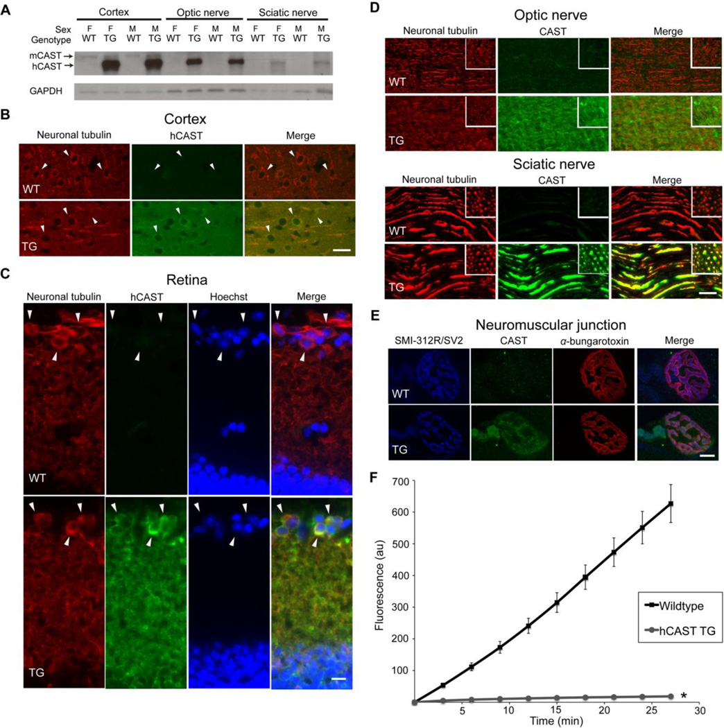
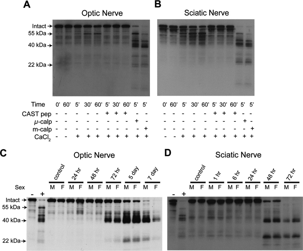
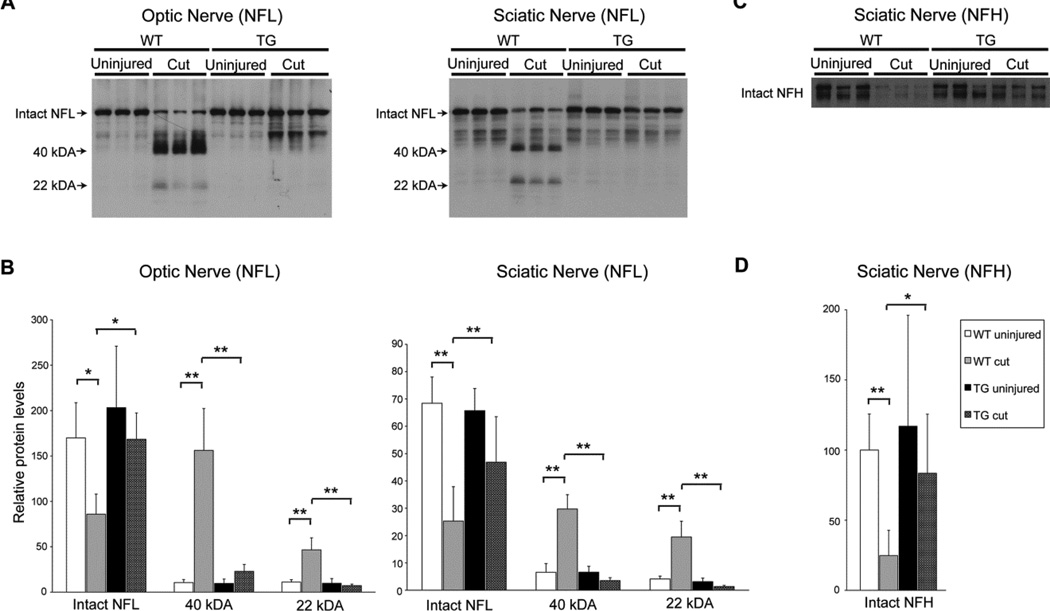
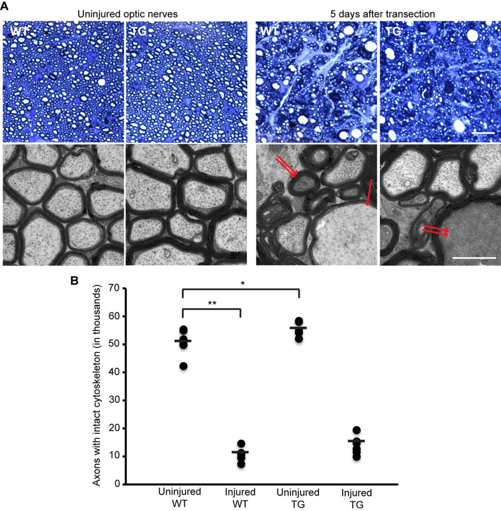
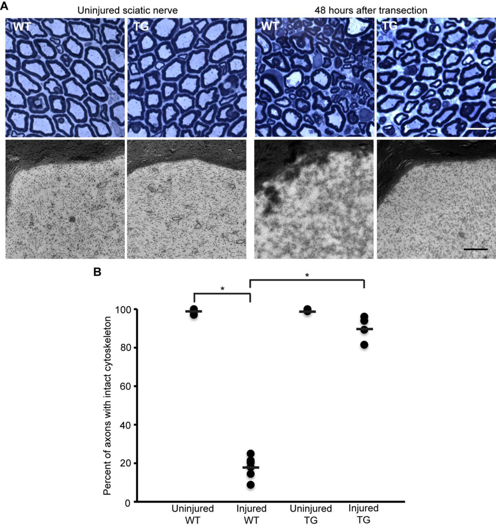
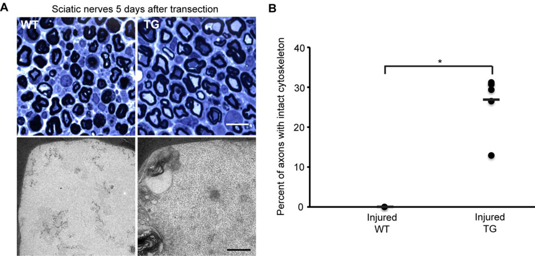
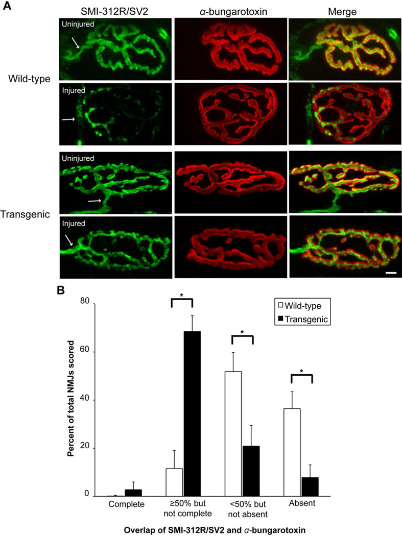
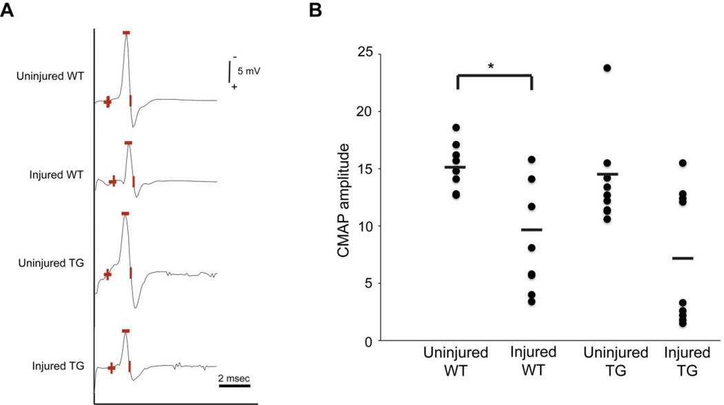
In both the central nervous system (CNS) and peripheral nervous system (PNS), transected axons undergo Wallerian degeneration. Even though Augustus Waller first described this process after transection of axons in 1850, the molecular mechanisms may be shared, at least in part, by many human diseases. Early pathology includes failure of synaptic transmission, target denervation, and granular disintegration of the axonal cytoskeleton (GDC). The Ca(2+)-dependent protease calpains have been implicated in GDC but causality has not been established. To test the hypothesis that calpains play a causal role in axonal and synaptic degeneration in vivo, we studied transgenic mice that express human calpastatin (hCAST), the endogenous calpain inhibitor, in optic and sciatic nerve axons. Five days after optic nerve transection and 48 h after sciatic nerve transection, robust neurofilament proteolysis observed in wild-type controls was reduced in hCAST transgenic mice. Protection of the axonal cytoskeleton in sciatic nerves of hCAST mice was nearly complete 48 h post-transection. In addition, hCAST expression preserved the morphological integrity of neuromuscular junctions. However, compound muscle action potential amplitudes after nerve transection were similar in wild-type and hCAST mice. These results, in total, provide direct evidence that calpains are responsible for the morphological degeneration of the axon and synapse during Wallerian degeneration.


Figures

Figure 1
Figure 1. Characterization of the human calpastatin transgenic mice
(A) Western blot of cortical and optic nerve homogenates and sciatic nerve supernatants (12 µg load) of human calpastatin (hCAST) transgenic (TG) mice and wild-type (WT) littermates was probed with antibodies targeting calpastatin (CAST; sc-20779) and glyceraldehyde 3-phosphate dehydrogenase (GAPDH). Calpastatin of mouse origin (mCAST) has a slightly slower mobility than hCAST (Schoch et al., in press). Other abbreviations: M, male; F, female. (B) Coronal sections of the cortex were immunostained for neuron-specific class III β-tubulin and hCAST (MAB3084). Cortical neurons are indicated by arrowheads. Scale bar=25 µm. (C) Transverse sections of retina were double immunolabeled with class III β-tubulin and hCAST antibodies and counterstained with nuclear Hoechst. Cells positive for the retinal ganglion cell marker, class III β-tubulin (Cui et al., 2003; Kwong et al., 2011), are indicated by arrowheads. Scale bar=10 µm. (D) Longitudinal and cross-sections (inset) of optic and sciatic nerves were immunostained for neuron-specific class III β-tubulin and CAST. Scale bar=25 µm. (E) Representative images of neuromuscular junctions (NMJs) from levator auris longus muscle stained with SMI-312R/SV2 antibodies (preterminal axons and the presynaptic compartment), α-bungarotoxin (postsynaptic membrane), and CAST antibody. Scale bar=10 µm. (D) Ex vivo analysis for functional hCAST in cortical homogenates from TG mice compared to WT littermates. When WT cortical homogenates (n=5) were added to the reaction mix, increased fluorescent signal from proteolysis of the calpain substrate Suc-Leu-Tyr-MNA was evident. However, the addition of TG cortical homogenates (n=5) robustly inhibited proteolysis of the fluorogenic substrate. Error bars represent SD. *p<0.0001 compared to WT. In TG mice, hCAST is functional and is present in the axonal compartment and synapse.
Figure 2
Figure 2. In vitro and in vivo proteolysis of neurofilament light
Homogenized optic nerves (A) and sciatic nerves (B) from wild-type (WT) mice were added to in vitro reactions containing a combination of calpastatin peptide (CAST pep), exogenous µ-calpain (µ-calp), exogenous m-calpain (m-calp), or CaCl2. Reactions were stopped at specified time points. Western blot was performed using an antiserum against full-length recombinant mouse neurofilament light (NFL). Calpain proteolysis resulted in loss of intact NFL, as well as generation of fragments with molecular weights of ~55, ~40, and ~22 kDa. For (C) and (D), WT mice underwent unilateral transection of optic nerves (C) and sciatic nerves (D) and were sacrificed at the indicated times. The two left-most lanes of each blot are controls in which optic or sciatic nerve homogenates underwent in vitro calpain digest. The “−” represents time 0 for the reaction in which CaCl2 was omitted, while “+” was stopped 5 minutes after adding CaCl2 and exogenous calpain. Western blot was performed using an antiserum against NFL. Other abbreviations: M, male; F, female. Both optic and sciatic nerve transection resulted in NFL proteolysis, which generated fragments with similar sizes (~40 and ~22 kDa) to those seen with the in vitro calpain digest. The latency period was longer after optic nerve transection.
Figure 3
Figure 3. In vivo neurofilament proteolysis in wild-type and transgenic mice
(A) Representative western blots of homogenates or supernatants of transected (cut) and contralateral uninjured optic and sciatic nerves from wild-type (WT) and transgenic (TG) mice using antiserum against neurofilament light (NFL). (B) Quantification of intact NFL and the ~40 and ~22 kDa proteolytic fragments. n=6–7 mice per group (optic nerve) and n=7–9 per group (sciatic nerve). (C) Representative western blot of supernatants of transected and contralateral uninjured sciatic nerves from WT and TG mice using an antibody targeting neurofilament heavy (NFH). NFH appears as a doublet, due to differential phosphorylation (Park et al., 2007). (D) Quantification of intact NFH doublet. Error bars represent SD. *p<0.01 **p<0.001. Human calpastatin blocked loss of full-length NFL and NFH, as well as the generation of the ~40 and ~22 kDa NFL fragments.
Figure 4
Figure 4. Axonal cytoskeleton 5 days after optic nerve transection in wild-type and transgenic mice
(A) Representative light and electron microscopic images of optic nerves from wild-type (WT) and transgenic (TG) mice. An optic nerve axon undergoing granular disintegration of the axonal cytoskeleton is marked by single arrow, while two of the axons undergoing “dark degeneration” are marked by double arrows. Scale bar=10 µm (light microscopy), 1 µm (electron microscopy). (B) Quantification of axons with intact cytoskeleton. n=6–7 mice per group. Each circle represents an individual nerve, while horizontal bars represent means. *p<0.017 **p<0.0001. Human calpastatin did not provide statistically significant morphological protection to transected optic nerve axons.
Figure 5
Figure 5. Axonal cytoskeleton 48 hours after sciatic nerve transection in wild-type and transgenic mice
(A) Representative light and electron microscopic images of sciatic nerves from wild-type (WT) and transgenic (TG) mice. Scale bar=10 µm (light microscopy), 0.5 µm (electron microscopy). (B) Percentage of axons scored as intact 48 hours after sciatic nerve transection (n=7 WT and 4 TG). Each circle represents an individual nerve, while horizontal bars represent means. *p<0.0001. Protection of the axonal cytoskeleton in sciatic nerves of TG mice was nearly complete at 48 hours post-transection.
Figure 6
Figure 6. Axonal cytoskeleton 5 days after sciatic nerve transection in wild-type and transgenic mice
(A) Representative light and electron microscopic images of sciatic nerves from wild-type (WT) and transgenic (TG) mice. Scale bar=10 µm (light microscopy), 0.5 µm (electron microscopy). (B) Percentage of axons scored as intact 5 days after sciatic nerve transection (n=7 WT and 6 TG). Each circle represents an individual nerve, while horizontal bars represent means. *p≤0.0002. Morphological protection of sciatic nerves was statistically significant 5 days post-transection with human calpastatin expression.
Figure 7
Figure 7. Denervation of neuromuscular junctions after sciatic nerve transection in wild-type and transgenic mice
(A) Representative images of neuromuscular junctions (NMJs) 18 hours after transection in wild-type (WT) and transgenic (TG) mice. SMI-312R/SV2 antibodies (green) label preterminal axons and the presynaptic compartment, while α-bungarotoxin (red) labels the postsynaptic membrane. Arrows mark the preterminal axon. Scale bar=10 µm. (B) NMJs were classified into one of 4 groups on the basis of SMI-312R/SV2 coverage of α-bungarotoxin labeling: (1) complete innervation, (2) ≥50% innervation but not complete, (3) <50% innervation, but not absent, (4) absent innervation. The Y axis represents the mean percentage of NMJs classified in each group (n=10 WT and 7 TG). Error bars represent SD. Data from the uninjured side is not shown. *p<0.0001. Human CAST expression preserved NMJ innervation after sciatic nerve transection.
Figure 8
Figure 8. Motor nerve responses after sciatic nerve transection in wild-type and transgenic mice
(A) Representative motor nerve responses from stimulation of wild-type (WT) and transgenic (TG) sciatic nerves 14 hours after transection and their uninjured contralateral controls. Amplitude was measured from baseline to peak (marked by the minus sign). (B) Compound muscle action potential (CMAP) amplitudes are plotted for 10 WT and 9 TG mice. Each circle represents an individual nerve, while horizontal bars represent means. *p<0.01. Human CAST expression does not improve motor nerve responses of transected sciatic nerves.

Similar articles

Cited by

References

    1. Ai J, Liu E, Wang J, Chen Y, Yu J, Baker AJ. Calpain inhibitor MDL-28170 reduces the functional and structural deterioration of corpus callosum following fluid percussion injury. J. Neurotrauma. 2007;24:960–978. - PubMed
    1. Bauder AR, Ferguson TA. Reproducible mouse sciatic nerve crush and subsequent assessment of regeneration by whole mount muscle analysis. J. Vis. Exp. 2012;60:e3606. - PMC - PubMed
    1. Beirowski B, Nógrádi A, Babetto E, Garcia-Alias G, Coleman MP. Mechanisms of axonal spheroid formation in central nervous system Wallerian degeneration. J. Neuropathol. Exp. Neurol. 2010;69:455–472. - PubMed
    1. Bignami A, Dahl D, Nguyen BT, Crosby CJ. The fate of axonal debris in Wallerian degeneration of rat optic and sciatic nerves. Electron microscopy and immunofluorescence studies with neurofilament antisera. J. Neuropathol. Exp. Neurol. 1981;40:537–550. - PubMed
    1. Blumbergs PC, Scott G, Manavis J, Wainwright H, Simpson DA, McLean AJ. Topography of axonal injury as defined by amyloid precursor protein and the sector scoring method in mild and severe closed head injury. J. Neurotrauma. 1995;12:565–572. - PubMed

Publication types

LinkOut - more resources