. 2013 May;45(5):556-62.
doi: 10.1038/ng.2602. Epub 2013 Mar 31.

Mutations in STAMBP, encoding a deubiquitinating enzyme, cause microcephaly-capillary malformation syndrome

Collaborators, Affiliations

Mutations in STAMBP, encoding a deubiquitinating enzyme, cause microcephaly-capillary malformation syndrome

Laura M McDonell et al. Nat Genet. 2013 May.

Abstract

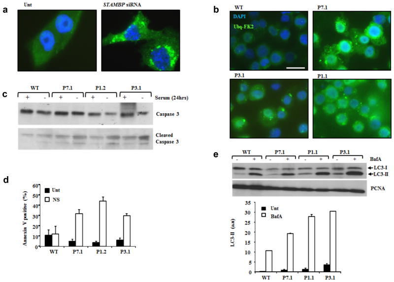
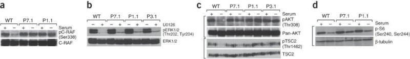
Microcephaly-capillary malformation (MIC-CAP) syndrome is characterized by severe microcephaly with progressive cortical atrophy, intractable epilepsy, profound developmental delay and multiple small capillary malformations on the skin. We used whole-exome sequencing of five patients with MIC-CAP syndrome and identified recessive mutations in STAMBP, a gene encoding the deubiquitinating (DUB) isopeptidase STAMBP (STAM-binding protein, also known as AMSH, associated molecule with the SH3 domain of STAM) that has a key role in cell surface receptor-mediated endocytosis and sorting. Patient cell lines showed reduced STAMBP expression associated with accumulation of ubiquitin-conjugated protein aggregates, elevated apoptosis and insensitive activation of the RAS-MAPK and PI3K-AKT-mTOR pathways. The latter cellular phenotype is notable considering the established connection between these pathways and their association with vascular and capillary malformations. Furthermore, our findings of a congenital human disorder caused by a defective DUB protein that functions in endocytosis implicates ubiquitin-conjugate aggregation and elevated apoptosis as factors potentially influencing the progressive neuronal loss underlying MIC-CAP syndrome.


Conflict of interest statement

COMPETING FINANCIAL INTERESTS

The authors declare no competing financial interests.

Figures

Figure 1
Figure 1
Neuroimaging and clinical features of MIC-CAP in Patient 9.1. T1-weighted sagittal (a) and axial (b) and T2-weighted coronal (c) images of the brain of Patient 9.1 at 3 months of age. Note the low-sloping forehead, simplified gyral pattern, increased extra-axial space, diffuse hypomyelination, and hippocampal hypoplasia. Photos of Patient 9.1 at 3 weeks (d) and 18 months (e) showing generalized capillary malformations of variable sizes and hypoplastic toenails (f).
Figure 2
Figure 2
Mutations in STAMBP cause MIC-CAP. (a) STAMBP (upper, chromosome 2, hg19: 74,056,114–74,094,295, RefSeq: NM_006463) and protein (lower, NP_006454.1) indicating MIC-CAP mutations. STAMBP contains a microtubule-interacting and transport (MIT) domain,, SH3 binding motif (SBM) (PX[V/I][D/N]RXXP), JAMM (JAB1/MPN/MOV34) motif, nuclear localization signal (NLS) and the distal ubiquitin recognition site (DUR). For c.279+5G>T in P2.1 (tissue from patient not available), a computational splicing model predicted the inclusion of an extra codon in exon 4 (p=1.9e-9, sign test). We validated this model using the known mutation in P7.1 (p=1.9e-9, sign test) (Supplementary Fig. 1a). Five out of six missense mutations are located in the MIT domain; required for the interaction of STAMBP with CHMP3, an ESCRT-III subunit. The sixth, Thr313Ile, located in the distal ubiquitin binding site within the JAMM domain, eliminates a hydrogen bond between the ubiquitin carbon backbone and STAMBP, likely decreasing ubiquitin binding to STAMBP (Supplementary Fig. 2). Two mutations were recurrent in multiple unrelated MIC-CAP families; Arg424* detected in Patients 3.1 and 4.1 and Arg38Cys detected in individuals P2.1, P7.1 and P8.1, suggestive of mutational hotspots in STAMBP. Within the ~5000 exomes in the NHLBI Exome variant server, only variant Arg38Cys was represented in 2 of 10756 alleles, suggesting a carrier frequency of approximately 1:5000 in a population of American/European ancestry, consistent with the prevalence of this very rare disorder. Western blot analysis of whole cell extracts (WCE) from LCLs P3.1, P5.1, P7.1, and P1.2, showing either equivocal (P3.1), reduced (P5.1) or absent STAMBP expression (P7.1, P1.2).
Figure 3
Figure 3
Elevated ubiquitin protein aggregates, apoptosis and autophagic flux in MIC-CAP. (a) Elevated conjugated-ubiquitin protein aggregates were observed following siRNA mediated silencing of STAMBP. T98G human medullablastoma cells were either untransfected (Unt) or transfected with siRNA against STAMBP. 24hrs post-transfection cells were stained with anti-FK2 and ubiquitin aggregates visualised by indirect immunofluorescence (IF). The extent of STAMBP knockdown is shown in Supplementary Figure 6b. (b) STAMBP-patient LCLs exhibit elevated levels of conjugated-ubiquitin protein aggregates. IF using anti-FK2 showed elevated levels of ubiquitinated protein aggregates in LCLs from P7.1, P3.1 and P1.1, compared to WT following 24hrs serum starvation. Scale bar represents 10μm. (c) STAMBP-patient LCLs exhibit elevated levels of apoptosis following 24hrs serum starvation. Elevated levels of cleaved caspase 3 were observed in LCLs from P7.1, P1.2 and P3.1, compared to WT, following serum starvation (24hrs). (d) Under similar conditions to (c), elevated levels of annexin V were observed in MIC-CAP patient LCLs P7.1, P1.2 and P3.1, compared to WT. Unt; untreated, NS: no-serum. Mean of four seperate determinations + sd. (e) Elevated autophagic flux, as demonstrated by LC3-II expression, was seem in multiple MIC-CAP LCLs, following treatment with Bafilomycin A (BafA; 100nM 2hrs), compared to WT LCLs. These data are consistent with elevated levels of autophagosomes in STAMBP-mutant patient LCLs compared to WT. Mean of three separate determinations ± sd.
Figure 4
Figure 4
Elevated RAS-GTP (active RAS) and activated PI3-kinase in MIC-CAP. (a) Schematic overview of core components of the RAS-MAPK and PI3K-AKT-mTOR networks highlighting this inter-connectivity. As well as interacting with the ESCRT machinery and STAM, STAMBP has been shown to interact with other important components of these signal transduction pathways including the Grb2 adaptor and the class II PI3-kinase catalytic subunit. (b) GTP-bound active RAS levels were precipitated from whole cell extracts using recombinant RAF1-RBD (RAS binding domain) GST-beads followed by Western blotting for RAS. GDP was shown to effectively compete any interaction as expected. Elevated levels of RAS-GTP were pulled down from P7.1 and P.1 MIC-CAP patient cells compared to WT LCLs. Image J based quantification (a.u. arbitrary units) of active RAS-GTP from three separate experiments are represented (± sd) in the graph. (c) Serum starvation (24hrs) reduced PI3-kinase activation in WT LCLs as monitored by phosphorylation of of the PI3K subunit p55-Y199 and p85-Y458. Phospho-PI3K levels were found to be elevated in extracts from P7.1 and P1.1 MIC-CAP LCLs either endogenously or following serum starvation suggestive of hyperactive and insensitive PI3K activity.
Figure 5
Figure 5
Elevated and insensitive RAS-MAPK and PI3K-AKT-mTOR signaling in MIC-CAP. (a) Serum starvation (24hrs) inhibits C-RAF activation (C-RAF-pS338) in wild-type (WT) LCLs in contrast to LCLs from P7.1 and P1.1. (b). LCLs were either treated (+) or untreated (-) with 10μM U0126, a specific MEK1/2 inhibitor for 1hr (Fig. 4a). Cells were harvested and WCEs were probed for phospho-ERK1/2 levels, which is mediated by MEK. Insensitivity to this treatment (as measured by relative phospho-ERK1/2 levels remaining after treatment with this MEK inhibitor), would reflect the magnitude/intensity of signal transduction from RAF to MEK to ERK (Fig. 4a). Residual pERK1/2 (pT202/pY204) signal (MEK-dependent phosphorylation) was seen in MIC-CAP LCLs, in contrast to WT. This phenotype is underscored following titration of U0126 in various MIC-CAP LCLs compared to WT (Supplementary Fig. 6e). Collectively, these data indicate a greater strength of MEK1/2 activity in MIC-CAP LCLs compared to WT cells. (c) Serum starvation (24hrs) reduces phosphorylation (activation) on AKT (T308) and on TSC2 at T1462, an AKT-dependent inhibitory TSC2 phosphorylation in WT LCLs in contrast to MIC-CAP LCLs from P7.1, P1.1 and P3.1. The TSC1/2 complex is the principal negative regulator of mTOR kinase complex (Fig. 4a). These data are consistent with active signal transduction from PI3K-AKT-mTOR in MIC-CAP cells under these conditions. (d) S6 protein is phosphorylated by S6-kinase in an mTOR-dependent fashion (Fig. 4a). Consistent with active signal transduction in this pathway under serum starvation conditions MIC-CAP LCLs from P7.1 and P1.1 maintained S6 phosphorylation on S240/S244 in contrast to WT LCLs.

References

    1. Carter MT, et al. A new syndrome with multiple capillary malformations, intractable seizures, and brain and limb anomalies. Am J Med Genet A. 2011;155:301–306. - PubMed
    1. Isidor B, Barbarot S, Bénéteau C, Le Caignec C, David A. Multiple capillary skin malformations, epilepsy, microcephaly, mental retardation, hypoplasia of the distal phalanges: Report of a new case and further delineation of a new syndrome. Am J Med Genet A. 2011;155:1458–1460. - PubMed
    1. Mirzaa GM, et al. The microcephaly-capillary malformation syndrome. Am J Med Genet A. 2011;155A:2080–2087. - PMC - PubMed
    1. Carter MT, Boycot KM. Microcephaly–capillary malformation syndrome: A story of rapid emergence of a new recognizable entity. Am J Med Genet A. 2011;155:2078–2079. - PubMed
    1. Jacobs AH, Walton RG. The incidence of birthmarks in the neonate. Pediatrics. 1976;58:218–222. - PubMed

Publication types

MeSH terms

Substances

LinkOut - more resources