

Capillary-free vascularized retina in patients with aggressive posterior retinopathy of prematurity and late retinal capillary formation
- PMID: 23542906
- PMCID: PMC3596613
- DOI: 10.3341/kjo.2013.27.2.109
Capillary-free vascularized retina in patients with aggressive posterior retinopathy of prematurity and late retinal capillary formation
Abstract
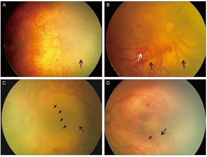
Purpose: To report the clinical features, clinical course, and treatment outcomes after laser photocoagulation in infants with aggressive posterior retinopathy of prematurity (APROP) and capillary-free zones in vascularized retina.
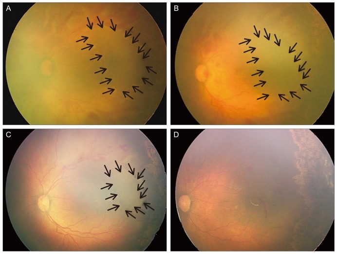
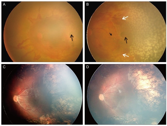
Methods: Six patients (12 eyes) with APROP and capillary-free zones in vascularized retina were retrospectively reviewed. Twelve eyes of six infants were included and were treated with laser photocoagulation for avascular retina and for capillary-free zones in vascularized retina, except for the posterior pole, and fundus findings were photographically-documented in sequence. In addition, anatomic and visual outcomes were evaluated with complications of APROP.
Results: Among all of the consecutive infants with APROP, capillary-free zones in vascularized retina were demonstrated in 24% of the infants. All of the infants were >27 weeks of gestation age and had birth weights >1,000 g. After laser treatment, 7 eyes (58.3%) had favorable outcomes, and late capillary filling in capillary-free zones of vascularized retina were noted, however 4 eyes (33.3%) progressed to retinal detachment and 1 eye (8.3%) was complicated by a retinal fold-distorting posterior pole. The visual outcomes were associated with anatomic outcomes.
Conclusions: The anatomic outcomes in infants with APROP who had capillary-free zones were comparable to previously reported infants with APROP. The late capillary filling of capillary-free zones in vascularized retina was noted, and angiogenesis was considered to be involved. This process toward normal capillary formation or neovascularization in APROP, might determine its outcome.
Keywords: Aggressive posterior retinopathy of prematurity; Angiogenesis; Capillaries.
Conflict of interest statement
No potential conflict of interest relevant to this article was reported.
Figures
References
-
- International Committee for the Classification of Retinopathy of Prematurity. The International Classification of Retinopathy of Prematurity revisited. Arch Ophthalmol. 2005;123:991–999. - PubMed
-
- Early Treatment for Retinopathy of Prematurity Cooperative Group. Revised indications for the treatment of retinopathy of prematurity: results of the early treatment for retinopathy of prematurity randomized trial. Arch Ophthalmol. 2003;121:1684–1694. - PubMed
-
- Morizane H. Initial sign and clinical course of the most severe form of acute proliferative retrolental fibroplasia (type II) Nihon Ganka Gakkai Zasshi. 1976;80:54–61. - PubMed
-
- Drenser KA, Trese MT, Capone A., Jr Aggressive posterior retinopathy of prematurity. Retina. 2010;30(4 Suppl):S37–S40. - PubMed
MeSH terms
LinkOut - more resources
Full Text Sources
Other Literature Sources
