. 2013 Aug;22(8):1750-7.
doi: 10.1007/s00586-013-2763-z. Epub 2013 Apr 1.

Clinical characteristics of intraspinal facet cysts following microsurgical bilateral decompression via a unilateral approach for treatment of degenerative lumbar disease

Affiliations

Clinical characteristics of intraspinal facet cysts following microsurgical bilateral decompression via a unilateral approach for treatment of degenerative lumbar disease

Minori Kato et al. Eur Spine J. 2013 Aug.

Abstract

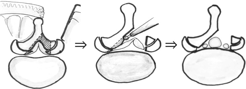
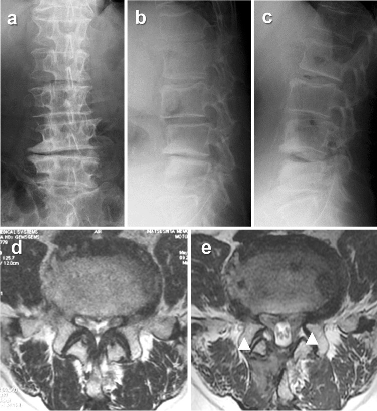
Purpose: Primary intraspinal facet cysts in the lumbar spine are uncommon, but it is unclear whether cyst incidence increases following decompression surgery and if these cysts negatively impact clinical outcome. We examined the prevalence, clinical characteristics, and the risk factors associated with intraspinal facet cysts after microsurgical bilateral decompression via a unilateral approach (MBDU).

Methods: We studied 230 patients treated using MBDU for lumbar degenerative disease (133 men and 97 women; mean age 70.3 years). Clinical status, as assessed by the Japanese Orthopedic Association (JOA) score and findings on X-ray and magnetic resonance images, was evaluated prior to surgery and at both 3 months and 1 year after surgery. The prevalence of intraspinal facet cysts was determined and preoperative risk factors were defined by comparing presurgical findings with clinical outcomes.

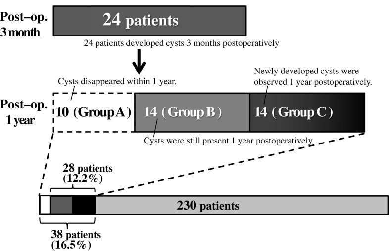
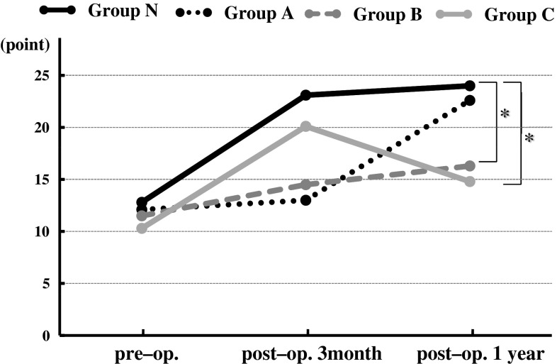
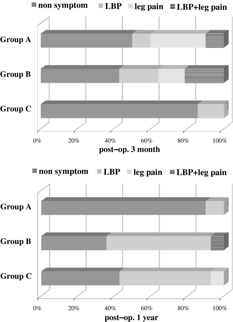
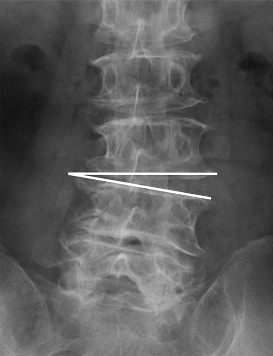
Results: Thirty-eight patients (16.5%) developed intraspinal facet cysts within 1 year postoperatively, and 24 exhibited cysts within 3 months. In 10 patients, the cysts resolved spontaneously 1 year postoperatively. In total, 28 patients (12.2%) had facet cysts 1 year postoperatively. The mean JOA score of patients with cysts 1 year postoperatively was significantly lower than that of patients without cysts. This poor clinical outcome resulted from low back pain that was not improved by conservative treatment. Most cases with spontaneous cyst disappearance were symptom-free 1 year later. The preoperative risk factors for postoperative intraspinal facet cyst formation were instability (OR 2.47, P = 0.26), scoliotic disc wedging (OR 2.23, P = 0.048), and sagittal imbalance (OR 2.22, P = 0.045).

Conclusions: Postoperative intraspinal facet cyst formation is a common cause of poor clinical outcome in patients treated using MBDU.


Figures

Fig. 1
Fig. 1
Schema showing our modified microsurgical bilateral decompression via a unilateral approach (MBDU) method (see “Materials and methods” and “Surgical procedure” for details)
Fig. 2
Fig. 2
Prevalence of intraspinal facet cysts. Thirty-eight patients (16.5 %) developed postoperative intraspinal facet cysts developed at some point during the first postoperative year. In 28 patients, the facet cysts persisted 1 year after surgery (12.2 %)
Fig. 3
Fig. 3
Graph showing clinical outcome according to the Japanese Orthopedic Association scoring system (JOA score). The asterisk indicates the recovery ratio of JOA score in Groups B and C were significantly lower than that in Group N (P < 0.05)
Fig. 4
Fig. 4
Graph showing the symptoms of patients with cysts. Three months postoperatively, about half of the patients in GroupsA and B had several symptoms. One year postoperatively, most patients in GroupA were symptom-free, while more than half of the patients in GroupsB and C had low back pain
Fig. 5
Fig. 5
Schema showing scoliotic disc wedging represented by the line. Preoperative scoliotic disc wedging was defined as wedging of >5° on anteroposterior X-rays in supine position
Fig. 6
Fig. 6
Representative case. A 68-year-old man with L4–5 degenerative lumbar spondylolisthesis and disc wedging reported intermitted claudication due to leg pain (JOA score of 13). After MBDU, intermitted claudication disappeared, but disabling lower back pain appeared 2 months postoperatively that resisted conservative treatment (1 year JOA score 15). ac Preoperative X-rays: a anteroposterior view, b lateral view (flexion), c lateral view (extension). d, e Axial T2-weighted magnetic resonance imaging of L4–5: d preoperative, e 1 year postoperatively (arrowheads indicate intraspinal facet cysts)

References

    1. Poletti CE. Central lumbar stenosis caused by ligamentum flavum: unilateral laminotomy for bilateral ligamentectomy: preliminary report of two cases. Neurosurgery. 1995;37(2):343–347. doi: 10.1227/00006123-199508000-00025. - DOI - PubMed
    1. McCulloch JA, Young PA. Essentials of spinal microsurgery. Philadelphia: Lippincott-Raven; 1998.
    1. Spetzger U, Bertalanffy H, Naujokat C, et al. Unilateral laminotomy for bilateral decompression of lumbar spinal stenosis. Part I: anatomical and surgical considerations. Acta Neurochir (Wien) 1997;139(5):392–396. doi: 10.1007/BF01808872. - DOI - PubMed
    1. Spetzger U, Bertalanffy H, Reinges MH, et al. Unilateral laminotomy for bilateral decompression of lumbar spinal stenosis. Part II: clinical experiences. Acta Neurochir (Wien) 1997;139(5):397–403. doi: 10.1007/BF01808874. - DOI - PubMed
    1. Weiner BK, Walker M, Brower RS, et al. Microdecompression for lumbar spinal canal stenosis. Spine. 1999;24(21):2268–2272. doi: 10.1097/00007632-199911010-00016. - DOI - PubMed

MeSH terms

LinkOut - more resources