. 2013 Jun;83(6):1237-46.
doi: 10.1124/mol.113.085068. Epub 2013 Mar 29.

A re-evaluation of the role of hCTR1, the human high-affinity copper transporter, in platinum-drug entry into human cells

Affiliations

A re-evaluation of the role of hCTR1, the human high-affinity copper transporter, in platinum-drug entry into human cells

Kristin D Ivy et al. Mol Pharmacol. 2013 Jun.

Abstract

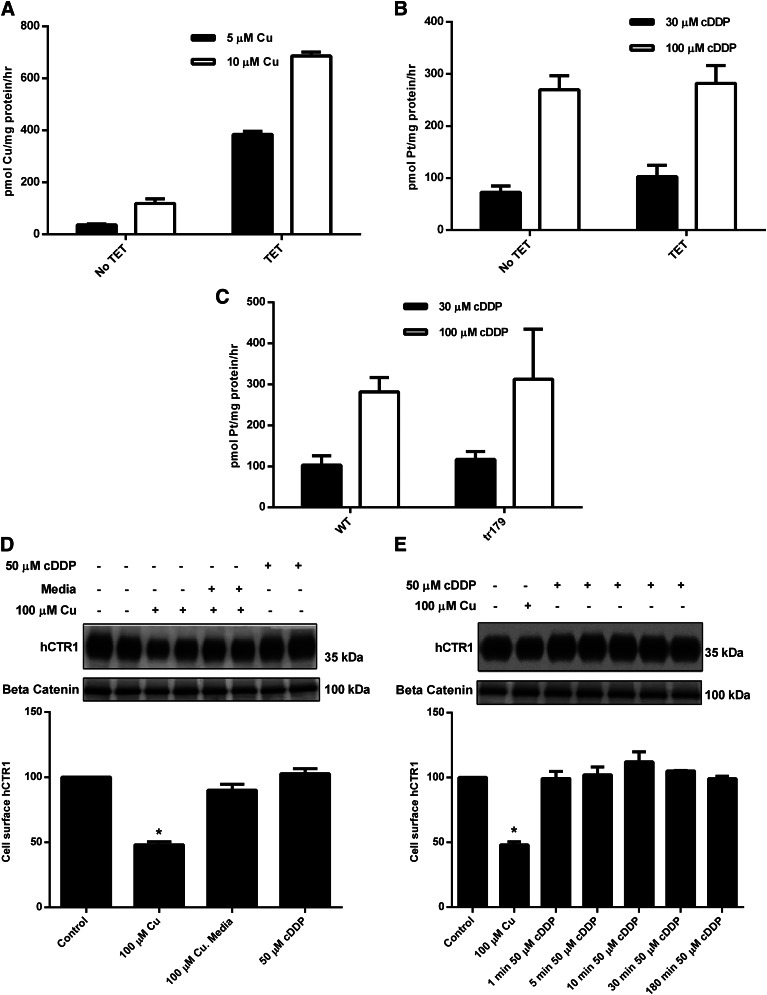
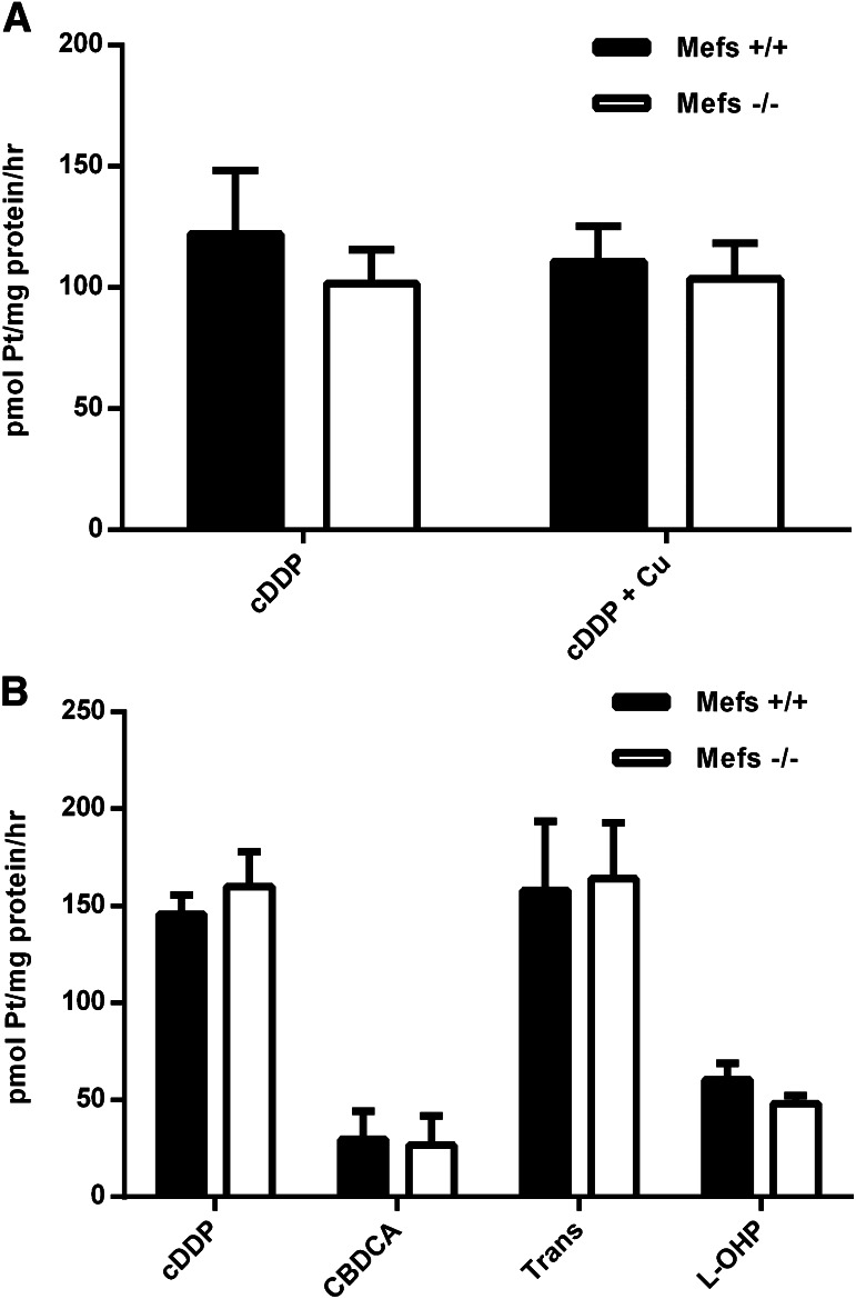
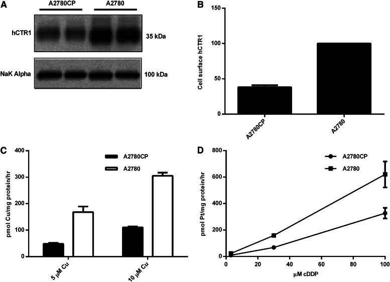
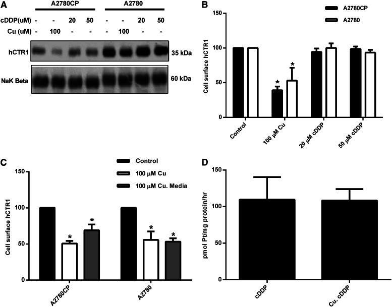
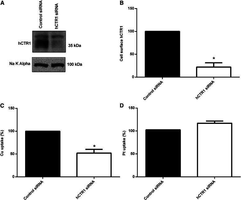
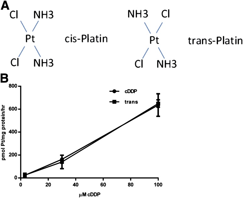
Cisplatin (cDDP) is an anticancer drug used in a number of malignancies, including testicular, ovarian, cervical, bladder, lung, head, and neck cancers. Its use is limited by the development of resistance, often rationalized via effects on cellular uptake. It has been claimed that human copper transporter 1 (hCTR1), the human high-affinity copper transporter, is the major entry pathway for cDDP and related drugs via a mechanism that mimics copper. This is an unexpected property of hCTR1, a highly selective copper (I) transporter. We compared the uptake rates of copper with cDDP (and several analogs) into human embryonic kidney 293 cells overexpressing wild-type or mutant hCTR1, mouse embryonic fibroblasts that do or do not express CTR1, and human ovarian tumor cells that are sensitive or resistant to cDDP. We have also compared the effects of extracellular copper, which causes regulatory endocytosis of hCTR1, to those of cDDP. We confirm the correlation between higher hCTR1 levels and higher platinum drug uptake in tumor cells sensitive to the drug. However, we show that hCTR1 is not the major entry route of platinum drugs, and that the copper transporter is not internalized in response to extracellular drug. Our data suggest the major entry pathway for platinum drugs is not saturable at relevant concentrations and not protein-mediated. Clinical trials have been initiated that depend upon regulating membrane levels of hCTR1. If reduced drug uptake is a major factor in resistance, hCTR1 is unlikely to be a productive target in attempts to enhance efficacy, although the proteins involved in copper homeostasis may play a role.


Figures

Fig. 1.
Fig. 1.
Copper uptake, cDDP uptake, and cell surface expression of hCTR1 in HEK293 cells. HEK 293 cells overexpressing hCTR1 were induced with tetracycline (TET) 48 hours prior to the experiment. (A) 64Cu uptake was measured in cells incubated with 5 or 10 µM copper for 30 minutes in media. (B and C) cDDP uptake was measured in cells incubated with 30 or 100 µM cDDP for 5 hours. (D) Tetracycline-induced cells were incubated with 100 µM copper for 30 minutes, and then washed three times with media and placed back in media for 30 minutes, or incubated with 50 µM cDDP for 30 minutes. (E) Tetracycline-induced HEK cells were incubated with 100 µM copper for 1 hour, or 50 µM cDDP for 1, 5, 10, 30, or 180 minutes. (D and E) Cells were biotinylated, and the biotinylated protein was analyzed on Western blots. hCTR1 protein was detected using an anti-FLAG antibody, and the protein levels were normalized to β-catenin, loading control. WT, wild type. *Statistical analysis was performed using Student's t test, and P < 0.05 was considered statically significant.
Fig. 2.
Fig. 2.
Platinum uptake rates in Mefs. (A) Mefs (+/+) and (−/−) were incubated with 30 µM cDDP or 30 µM cDDP and 100 µM copper for 5 hours. (B) Mefs (+/+) and (−/−) were incubated with 30 µM cDDP, carboplatin (CBDCA), transplatin (Trans), or oxaliplatin (L-OHP) for 5 hours.
Fig. 3.
Fig. 3.
hCTR1 protein expression, copper uptake, and cDDP uptake in human ovarian carcinoma cells. (A) cDDP-resistant (A2780CP) and cDDP-sensitive (A2780) cells were fractionated, and plasma membranes were obtained and analyzed on Western blots. hCTR1 was detected using an anti-CTR1 antibody (rabbit). The Na+,K+-ATPase α subunit was the loading control. (B) Biotinylated hCTR1 protein was quantified using Quality One software (Bio-Rad), and the protein levels were normalized to the Na+,K+-ATPase α subunit. (C) 64Cu uptake was measured in cells incubated with 5 or 10 µM copper for 30 minutes in media. (D) cDDP uptake was measured in cells incubated with 3, 30, or 100 µM cDDP for 5 hours in media.
Fig. 4.
Fig. 4.
Surface expression of hCTR1 and the effects of copper and cDDP. (A) cDDP-resistant (A2780CP) and cDDP-sensitive (A2780) cells were incubated with 100 µM copper, 20 µM cDDP, or 50 µM cDDP for 1 hour. (C) Cells were incubated with 100 µM copper for 30 minutes or 100 µM Cu and then washed three times with media and placed back in media for 30 minutes. (A and C) Cells were biotinylated, and the biotinylated protein was analyzed on Western blots using Quality One software. hCTR1 protein was detected using an anti-hCTR1(rabbit) antibody. The Na+,K+-ATPase β subunit was used as the loading control. (B) Western blot quantitation of (A). (D) A2780 cells were incubated with 100 µM copper for 1 hour, rinsed three times with media, and then placed back in fresh media with 30 µM cDDP for 3 hours. *Statistical analysis was performed using Student's t test, and P < 0.05 was considered statically significant.
Fig. 5.
Fig. 5.
hCTR1 knockdown in A2780 cells. (A) cDDP-sensitive (A2780) cells were transiently transfected with siRNA duplexes against CTR1 or siRNA negative control in Opti-MEM medium. Cells were biotinylated, and the biotinylated protein was analyzed on Western blots. hCTR1 protein was detected using an anti-CTR1 antibody (rabbit). (B) Biotinylated hCTR1 protein was quantified using Quality One software (Bio-Rad), and the protein levels were normalized to the Na+,K+-ATPase α subunit. After transient transfection, cells were incubated with 5 µM copper for 1 hour (C) or 30 µM cDDP for 3 hours (D). In (C), Cu uptake is expressed as a percentage of control (control rate 129.01 pmol of Cu/mg of protein/h), and in (D), cDDP uptake is expressed as a percentage of control (control rate 92.05 pmol of Pt/mg of protein/h). *Statistical analysis was performed using Student's t test, and P < 0.05 was considered statically significant.
Fig. 6.
Fig. 6.
Cisplatin versus transplatin (trans) cDDP uptake. (A) The chemical structure of cisplatin and transplatin are shown. (B) cDDP- sensitive (A2780) cells were incubated with 3, 30, or 100 μM cDDP or transplatin for 5 hours. Platinum content was measured by inductively coupled plasma mass spectrometry and is shown as pmol of Pt/mg of protein/h.

References

    1. Ahmed Z, Deyama Y, Yoshimura Y, Suzuki K. (2009) Cisplatin sensitivity of oral squamous carcinoma cells is regulated by Na+,K+-ATPase activity rather than copper-transporting P-type ATPases, ATP7A and ATP7B. Cancer Chemother Pharmacol 63:643–650 - PubMed
    1. Andrews PA, Howell SB. (1990) Cellular pharmacology of cisplatin: perspectives on mechanisms of acquired resistance. Cancer Cells 2:35–43 - PubMed
    1. Andrews PA, Velury S, Mann SC, Howell SB. (1988) cis-Diamminedichloroplatinum(II) accumulation in sensitive and resistant human ovarian carcinoma cells. Cancer Res 48:68–73 - PubMed
    1. Ashino T, Sudhahar V, Urao N, Oshikawa J, Chen GF, Wang H, Huo Y, Finney L, Vogt S, McKinney RD, et al. (2010) Unexpected role of the copper transporter ATP7A in PDGF-induced vascular smooth muscle cell migration. Circ Res 107:787–799 - PMC - PubMed
    1. Beretta GL, Gatti L, Tinelli S, Corna E, Colangelo D, Zunino F, Perego P. (2004) Cellular pharmacology of cisplatin in relation to the expression of human copper transporter CTR1 in different pairs of cisplatin-sensitive and -resistant cells. Biochem Pharmacol 68:283–291 - PubMed

Publication types