. 2013 May 1;190(9):4483-7.
doi: 10.4049/jimmunol.1300212. Epub 2013 Mar 29.

Cutting Edge: memory regulatory t cells require IL-7 and not IL-2 for their maintenance in peripheral tissues

Affiliations

Cutting Edge: memory regulatory t cells require IL-7 and not IL-2 for their maintenance in peripheral tissues

Iris K Gratz et al. J Immunol. .

Abstract

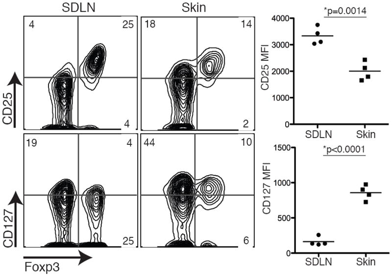
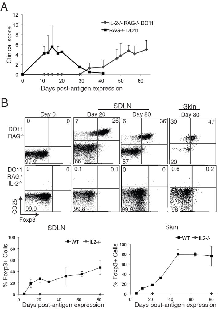
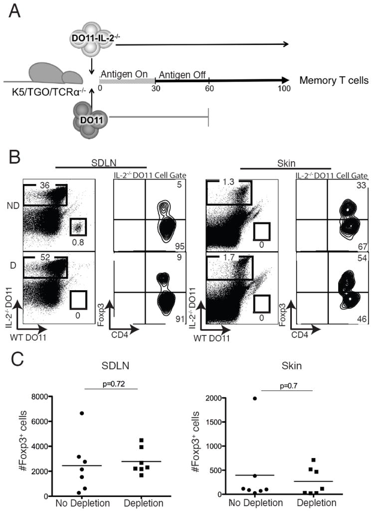
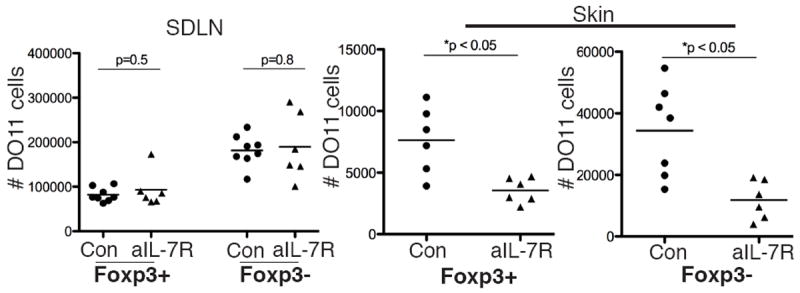
Thymic Foxp3-expressing regulatory T cells are activated by peripheral self-antigen to increase their suppressive function, and a fraction of these cells survive as memory regulatory T cells (mTregs). mTregs persist in nonlymphoid tissue after cessation of Ag expression and have enhanced capacity to suppress tissue-specific autoimmunity. In this study, we show that murine mTregs express specific effector memory T cell markers and localize preferentially to hair follicles in skin. Memory Tregs express high levels of both IL-2Rα and IL-7Rα. Using a genetic-deletion approach, we show that IL-2 is required to generate mTregs from naive CD4(+) T cell precursors in vivo. However, IL-2 is not required to maintain these cells in the skin and skin-draining lymph nodes. Conversely, IL-7 is essential for maintaining mTregs in skin in the steady state. These results elucidate the fundamental biology of mTregs and show that IL-7 plays an important role in their survival in skin.


Figures

Figure 1
Figure 1. Memory Tregs have reduced IL-2Rα expression and increased IL-7Rα expression
CD25 and CD127 expression was examined on mTregs isolated from the skin and SDLNs of K5/TGO/DO11 mice. Flow cytometry plots are gated on live CD4+DO11+ cells and column graphs are gated on live CD4+Foxp3+DO11+ cells. MFI, mean fluorescent intensity. Results are representative of 4 replicate experiments with > 3 mice per group.
Figure 2
Figure 2. IL-2 is required to generate pTregs in vivo
(A) Lymph node cells from DO11/Rag-/- or DO11/Rag-/-/IL-2-/- mice were adoptively transferred into K5/TGO/TCRα-/- hosts, Ova expression was induced in the skin, and severity of clinical skin disease measured. (B) Skin and SDLNs were harvested at specific times after antigen induction and DO11 cells analyzed by flow cytometry. Live CD4+DO11+ cells are shown. Error bars in 2A represent range. Results are representative of 2 replicate experiments with > 2 mice per group.
Figure 3
Figure 3. IL-2 is not required to maintain mTregs in the periphery
(A) To provide IL-2 in trans, WT CD90.1+DO11/Rag-/- cells (“DO11”) were co-transferred with CD90.2+DO11/Rag-/-/IL-2-/- cells (“DO11-IL-2-/-”) into K5/TGO/TCRα-/- hosts and Ova expression was induced in the skin for 30 days. Antigen was subsequently extinguished and 30 days later (i.e., day 60), WT DO11 cells were depleted or not depleted in control animals. DO11 memory cells were quantified 40 days after depletion (i.e., day 100). (B) Flow cytometry of DO11 cells harvested from skin and SDLNs at day 100 in mice where WT DO11 cells had been depleted (D) or not depleted (ND). (C) Absolute numbers of DO11-IL-2-/- cells expressing Foxp3 in the SDLN and skin in depleted and non-depleted mice on day 100. p = p value from 2-tailed unpaired t test. Results are pooled data from 3 replicate experiments with > 2 mice per group.
Figure 4
Figure 4. IL-7 is required to maintain mTregs in skin
Memory Tregs were generated by inducing Ova expression in the skin of K5/TGO/DO11 mice for 30 days and subsequently turning off antigen for > 30 days. Mice were subsequently treated with anti-IL-7Rα blocking antibody or control antibody for 21-28 days. Absolute number of Foxp3+ and Foxp3- DO11 cells in the skin and SDLNs after treatment with anti-IL-7Rα blocking antibody or control antibody are shown. p value from 2-tailed unpaired t test. Results are pooled data from 2 of 3 replicate experiments with > 3 mice per group.

References

    1. Bennett CL, Christie J, Ramsdell F, Brunkow ME, Ferguson PJ, Whitesell L, Kelly TE, Saulsbury FT, Chance PF, Ochs HD. The immune dysregulation, polyendocrinopathy, enteropathy, X-linked syndrome (IPEX) is caused by mutations of FOXP3. Nat Genet. 2001;27:20–21. - PubMed
    1. Fontenot JD, Gavin MA, Rudensky AY. Foxp3 programs the development and function of CD4+CD25+ regulatory T cells. Nat Immunol. 2003;4:330–336. - PubMed
    1. Rosenblum MD, Gratz IK, Paw JS, Lee K, Marshak-Rothstein A, Abbas AK. Response to self antigen imprints regulatory memory in tissues. Nature. 2011;480:538–542. - PMC - PubMed
    1. Rowe JH, Ertelt JM, Xin L, Way SS. Pregnancy imprints regulatory memory that sustains anergy to fetal antigen. Nature. 2012;490:102–106. - PMC - PubMed
    1. Miyara M, Yoshioka Y, Kitoh A, Shima T, Wing K, Niwa A, Parizot C, Taflin C, Heike T, Valeyre D, Mathian A, Nakahata T, Yamaguchi T, Nomura T, Ono M, Amoura Z, Gorochov G, Sakaguchi S. Functional delineation and differentiation dynamics of human CD4+ T cells expressing the FoxP3 transcription factor. Immunity. 2009;30:899–911. - PubMed

Publication types

MeSH terms

Substances