. 2013;8(3):e59759.
doi: 10.1371/journal.pone.0059759. Epub 2013 Mar 27.

Light-emitting channelrhodopsins for combined optogenetic and chemical-genetic control of neurons

Affiliations

Light-emitting channelrhodopsins for combined optogenetic and chemical-genetic control of neurons

Ken Berglund et al. PLoS One. 2013.

Abstract

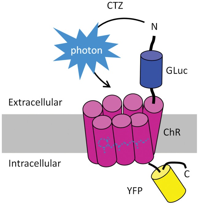
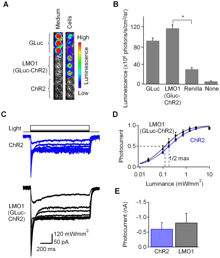
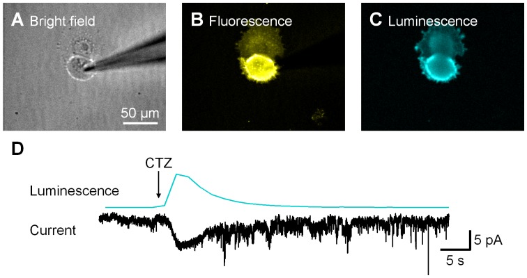
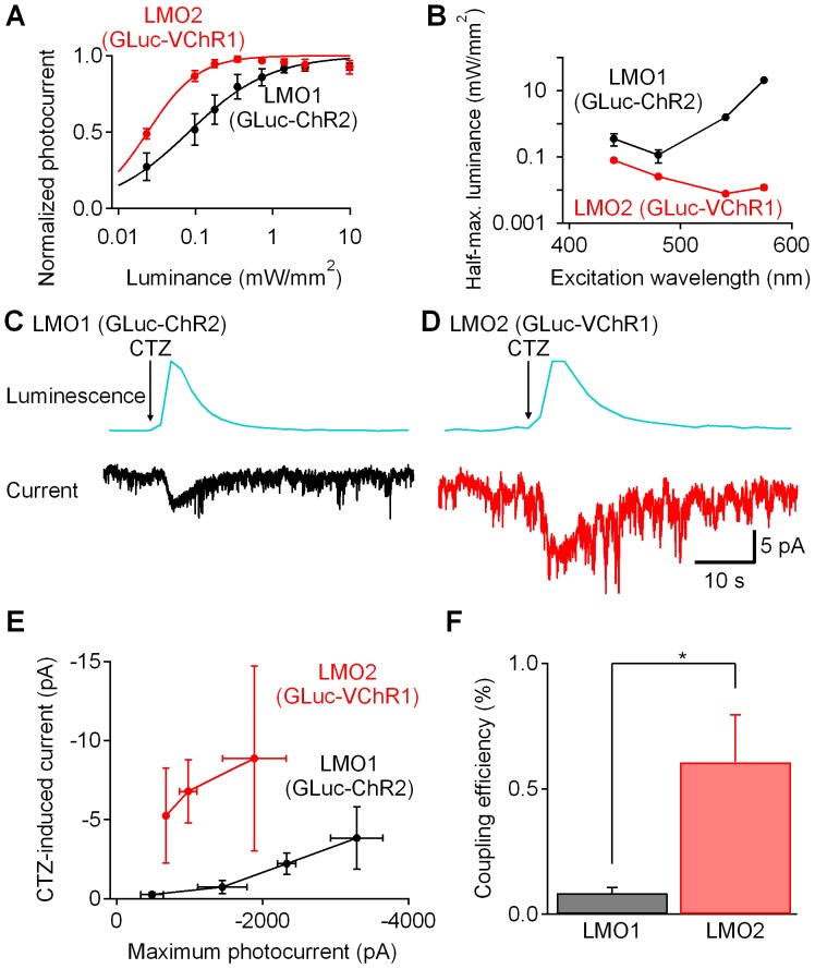
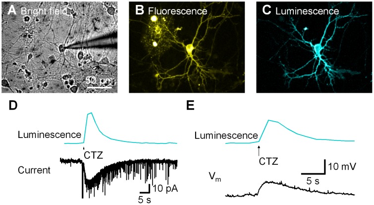
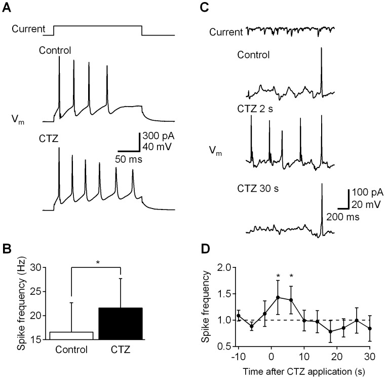
Manipulation of neuronal activity through genetically targeted actuator molecules is a powerful approach for studying information flow in the brain. In these approaches the genetically targeted component, a receptor or a channel, is activated either by a small molecule (chemical genetics) or by light from a physical source (optogenetics). We developed a hybrid technology that allows control of the same neurons by both optogenetic and chemical genetic means. The approach is based on engineered chimeric fusions of a light-generating protein (luciferase) to a light-activated ion channel (channelrhodopsin). Ionic currents then can be activated by bioluminescence upon activation of luciferase by its substrate, coelenterazine (CTZ), as well as by external light. In cell lines, expression of the fusion of Gaussia luciferase to Channelrhodopsin-2 yielded photocurrents in response to CTZ. Larger photocurrents were produced by fusing the luciferase to Volvox Channelrhodopsin-1. This version allowed chemical modulation of neuronal activity when expressed in cultured neurons: CTZ treatment shifted neuronal responses to injected currents and sensitized neurons to fire action potentials in response to subthreshold synaptic inputs. These luminescent channelrhodopsins--or luminopsins--preserve the advantages of light-activated ion channels, while extending their capabilities. Our proof-of-principle results suggest that this novel class of tools can be improved and extended in numerous ways.


Conflict of interest statement

Competing Interests: The authors have declared that no competing interests exist.

Figures

Figure 1
Figure 1. Design of luminopsins, luciferase-channelrhodopsin fusion proteins.
Gaussia luciferase (GLuc) is fused to the N-terminus of channelrhodopsin (ChR). Yellow fluorescent protein (YFP) is fused to the C-terminus of ChR. Application of the GLuc substrate coelenterazine (CTZ) leads to an enzymatic reaction resulting in the production of light (photons) and opening of the channel.
Figure 2
Figure 2. Preserved functionality of individual moieties within luminopsin-1.
(A) HEK cells were transfected with native, secreted Gaussia luciferase (GLuc), luminopsin-1 (LMO1; GLuc-ChR2 fusion gene), or with ChR2 alone and bioluminescence was determined by adding CTZ to the medium or to the cells. Only secreted GLuc produced signal in the medium, while bioluminescence generated by LMO1 was found only in cells. (B) Comparison of the bioluminescence signals obtained from cells (104 cells per well, 4 wells per group) transfected with native, secreted GLuc (GLuc), luminopsin-1 (LMO1, GLuc-ChR2 fusion construct), and Renilla luciferase (Renilla) as well as untransfected cells (None). Bioluminescence was comparable between native GLuc and GLuc within LMO1. Luminescence from LMO1 and Renilla was significantly different (*p<0.05; one-way ANOVA followed by Tukey’s test). (C) Comparison of photocurrents induced by physical light in HEK cells transfected with ChR2 (upper panel) and LMO1 (GLuc-ChR2; lower panel). (D) Luminance-photocurrents curves for ChR2 and LMO1 (GLuc-ChR2). When normalized, they showed similar half-maximal luminances (1/2 max). n = 3. (E) No significant differences in maximum photocurrents between ChR2 and LMO1 (GLuc-ChR2) were observed (p>0.05; two-tailed Students’ T-test; n = 3 each). Error bars denote S.E.M in this and subsequent figures.
Figure 3
Figure 3. Luminescence activated photocurrent.
LMO1 (GLuc-ChR2)-expressing HEK cell (A) was identified by YFP fluorescence (B) and patch-clamped. Coelenterazine (CTZ) application near the cell elicited bioluminescence (C). (D) Luminescence (upper trace) and luminescence-induced photocurrent (lower trace) were recorded simultaneously from the same cell.
Figure 4
Figure 4. Comparison between luminopsins LMO1 (GLuc-ChR2) and LMO2 (GLuc-VChR1).
(A) HEK cells were transfected with LMO1 (GLuc-ChR2) or LMO2 (GLuc-VChR1) (n = 4 and 5, respectively). Photocurrents to various intensities of 470 nm light were recorded and normalized to the maximum to compare light sensitivity between VChR1 and ChR2. The luminance-photocurrent curve of VChR1 is shifted to the left, indicating higher sensitivity of VChR1 to 470 nm light. (B) Half-maximum luminance was lowest at 470 nm for ChR2, and at 540 nm for VChR1, confirming red-shifted excitation spectrum of VChR1. VChR1 showed lower half-maximum luminances at all the wavelengths tested, indicating superior light sensitivity compared to ChR2 (n = 6 for LMO1 and 6 for LMO2). (C and D) CTZ application resulted in photocurrents in cells expressing LMO1 (GLuc-ChR2) (C) and LMO2 (GLuc-VChR1) (D). LMO2 (GLuc-VChR1) showed bigger photocurrent. (E) Correlation between maximum photocurrent induced by an arc lamp and luminescence-induced photocurrent from LMO1 (GLuc-ChR2) and LMO2 (GLuc-VChR1) expressing cells. n = 3 for each point. (F) Coupling efficiency of LMO2 (VChR1-GLuc) was significantly higher than that of LMO1 (ChR2-GLuc) (*p<0.05; two-tailed Students’ T-test; n = 12 and 9, respectively).
Figure 5
Figure 5. Luminescence-evoked responses in neurons.
Hippocampal neurons were transfected with LMO2 (GLuc-VChR1) and patch-clamped (A). Expression of the fusion protein was visualized by YFP fluorescence (B). Upon CTZ application, GLuc generated bioluminescence throughout the processes (C). This bioluminescence evoked both inward current under voltage-clamp (D) and depolarization under current-clamp (E).
Figure 6
Figure 6. Modulation of neuronal activity by luminescence.
Hippocampal neurons were transfected with LMO2 (GLuc-VChR1) and their intrinsic excitability was examined before and after CTZ application. (A) A square pulse (150 pA; top) was injected to a hippocampal neuron under current clamp, eliciting action potential firing (middle). After CTZ application, the neuron fired more action potentials (bottom). (B) Spike frequency with 150-pA current injection was significantly increased by CTZ (*p<0.05; two-tailed paired Students’ T-test; n = 3). (C) Prerecorded subthreshold spontaneous excitatory postsynaptic currents (sEPSCs; top) were injected into a neuron under current clamp, inducing single action potential (control; second from top). After CTZ application action potential firing was increased (CTZ 2 s; third from top) and returned to the baseline (CTZ 30 s; bottom). (D) On average, CTZ application transiently enhanced action potential firing up to 50%; n = 7.

References

    1. Luo L, Callaway EM, Svoboda K (2008) Genetic dissection of neural circuits. Neuron 57: 634–660. - PMC - PubMed
    1. Rogan SC, Roth BL (2011) Remote control of neuronal signaling. Pharmacol Rev 63: 291–315. - PMC - PubMed
    1. Magnus CJ, Lee PH, Atasoy D, Su HH, Looger LL, et al. (2011) Chemical and genetic engineering of selective ion channel-ligand interactions. Science 333: 1292–1296. - PMC - PubMed
    1. Miesenbock G (2011) Optogenetic Control of Cells and Circuits. Annu Rev Cell Dev Biol 27: 30.1–30.28. - PMC - PubMed
    1. Yizhar O, Fenno LE, Davidson TJ, Mogri M, Deisseroth K (2011) Optogenetics in neural systems. Neuron 71: 9–34. - PubMed

Publication types