. 2013;8(3):e60152.
doi: 10.1371/journal.pone.0060152. Epub 2013 Mar 27.

Inhibition of PIKfyve by YM-201636 dysregulates autophagy and leads to apoptosis-independent neuronal cell death

Affiliations

Inhibition of PIKfyve by YM-201636 dysregulates autophagy and leads to apoptosis-independent neuronal cell death

Sally Martin et al. PLoS One. 2013.

Abstract

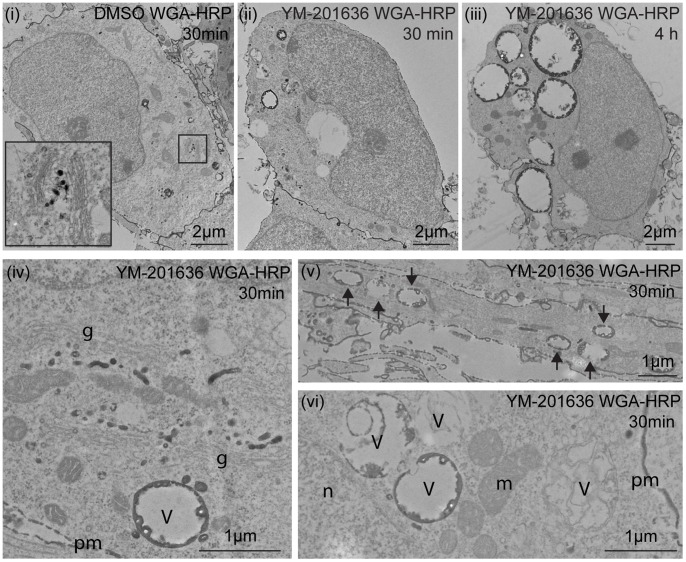
The lipid phosphatidylinositol 3,5-bisphosphate (PtdIns(3,5)P 2), synthesised by PIKfyve, regulates a number of intracellular membrane trafficking pathways. Genetic alteration of the PIKfyve complex, leading to even a mild reduction in PtdIns(3,5)P 2, results in marked neurodegeneration via an uncharacterised mechanism. In the present study we have shown that selectively inhibiting PIKfyve activity, using YM-201636, significantly reduces the survival of primary mouse hippocampal neurons in culture. YM-201636 treatment promoted vacuolation of endolysosomal membranes followed by apoptosis-independent cell death. Many vacuoles contained intravacuolar membranes and inclusions reminiscent of autolysosomes. Accordingly, YM-201636 treatment increased the level of the autophagosomal marker protein LC3-II, an effect that was potentiated by inhibition of lysosomal proteases, suggesting that alterations in autophagy could be a contributing factor to neuronal cell death.


Conflict of interest statement

Competing Interests: The authors would like to state that Sally Martin and Frederic A. Meunier are PLOS ONE Editorial Board members. This does not alter the authors’ adherence to all the PLOS ONE policies on sharing data and materials.

Figures

Figure 1
Figure 1. YM-201636 promotes an apoptosis-independent cell death in cultured primary hippocampal neurons.
(A) Primary hippocampal neurons were treated for 24 h with DMSO or 1 µM YM-201636 with and without 30 µM Z-VAD-fmk and imaged by brightfield microscopy. (B) Quantitation of neuronal survival following 24 h treatment with DMSO or 1 µM YM-201636 with and without 30 µM Z-VAD-fmk (ZVF), n = 12 fields of cells, 3 independent experiments. (C) Primary hippocampal neurons were treated for 4 h or 18 h with DMSO, 1 µM YM-201636 or 500 nM staurosporine and immunoblotted for cleaved caspase-3 and β-actin. (D) Primary hippocampal neurons were treated for 4 h with DMSO, 500 nM staurosporine with and without 30 µM ZVF, 30 µM ZVF or 1 µM YM-201636 and immunoblotted for cleaved caspase-3 and β-actin. (E-H) Primary hippocampal neurons were imaged for 48 h in real time using brightfield microscopy and analysed for total neurite length (E) and neurite length per neuron (F) (n = 20 fields in total from 4 wells of cells) or the percentage of dead (G) or vacuolated (H) cells (n = 4 wells). All results show mean ± SEM. Circle = DMSO, Square = YM-201636. The level of significance is shown relative to DMSO, *p<0.05, **p<0.01, ***p<0.001.
Figure 2
Figure 2. YM-201636 promotes vacuolation and endosomal compartments in hippocampal neurons.
(A) Primary hippocampal neurons were treated with DMSO or 1 µM YM-201636 for 4 h and processed for electron microscopy. Images show neuronal cell bodies at the level of the nucleus, demonstrating vacuolation in the presence of YM-201636. Grid size = 1 µm square. (B-D) Electron microscopic analysis of the vacuole area as a percentage of total cytoplasmic area (B) and the number of vacuoles per cell (C) shows a significant increase in vacuole size and number following 4 h treatment with 1 µM YM-201636, **p<0.01, ***p<0.001. (D) Histogram of number of vacuoles relative to their size (representative experiment), n = 12 (DMSO) or 11 (YM-201636) cells. (E) Examples of vacuole phenotypes detected in primary hippocampal neurons treated with 1 µM YM-201636 for 4 h. (F) Primary hippocampal neurons were treated with DMSO or 1 µM YM-201636 for 4 h or 18 h and immunolabelled for LAMP1, EEA1 and ß3-tubulin. 3D projections are shown. Scale bar = 10 µm.
Figure 3
Figure 3. The effect of YM-201636 on endosomal and retrograde trafficking in hippocampal neurons.
(A) Primary hippocampal neurons were treated with DMSO or 1 µM YM-201636 for 2 h then supplemented with 1 µg/ml CTB-Alexa555 for a further 2 h. Cells were fixed and immunolabelled for GM130. Representative 3D projections are shown. (B) The integrated intensity (per µm2) of CTB-Alexa555 in Golgi complex, as defined by GM130, the integrated intensity of GM130 and the size of the area analysed was measured and the percentage change between conditions determined. (C) The change in total CTB-Alexa555 integrated intensity within the cell body and the area (µm2) of the cell body was determined. (mean ± SEM, n = 3 independent experiments, 11–20 cells per experiment). Significances relative to DMSO *p<0.05, **p<0.01, ***p<0.001 (D) Primary hippocampal neurons were treated with DMSO or 1 µM YM-201636 for 3.5 h then supplemented with 25 µg/ml transferrin-Alexa555 (Tf-Alexa555) for a further 30 min. Cells were fixed and immunolabelled for LAMP1. Scale bar = 10 µm.
Figure 4
Figure 4. Effect of YM-201636 on WGA trafficking by immunocytochemistry.
Primary hippocampal neurons were treated with DMSO or 1 µM YM-201636 for 4 h, then supplemented with 5 µg/ml WGA-Alexa555 for the final 5 min (A,B) or 30 min (C,D). Cells were fixed, immunolabelled for EEA1 and LAMP1, and imaged by confocal microscopy. (B,D) The intensity of WGA-Alexa555 per µm2 in the cell body and the total area of the cell body were determined in the YM-201636–treated cells relative to DMSO. (E,F) Primary hippocampal neurons were treated with DMSO or 1 µM YM-201636 in the presence of 5 µg/ml WGA-Alexa555 for the full 4 h. Cells were fixed, immunolabelled for GM130 and imaged by confocal microscopy. (F) The amount of WGA-Alexa555 fluorescent intensity in the cell body/µm2 of the YM-2016363 treated cells was determined relative to DMSO. The area of the cell bodies analysed was also determined (mean ± SEM, n = 3 independent experiments, 9–28 cells per experiment). Significances relative to DMSO **p<0.01. Scale bar = 10 µm.
Figure 5
Figure 5. Ultrastructural analysis of the effect of YM-201636 on WGA trafficking.
Primary hippocampal neurons were treated with (i) DMSO for 3.5 h and 10 µg/ml WGA-HRP for a further 30 min, (ii, iv–vi) 1 µM YM-201636 for 3.5 h and 10 µg/ml WGA-HRP for a further 30 min or (iii) 1 µM YM-201636 and 10 µg/ml WGA-HRP for 4 h. Cells were fixed, processed for DAB cytochemistry and imaged by electron microscopy. (i) In DMSO-treated cells WGA was identified in small vacuoles and vesicles. (ii) When WGA was added following inhibition of PIKfyve for 3.5 h, it was observed in a subset (∼45%) of large vacuoles. (iii) When WGA was continually present during the inhibition of PIKfyve, most (∼90%) of vacuoles contained DAB reaction product. (iv) Transport of WGA-HRP to the peri-Golgi region was unperturbed by treatment with YM-201636. (v) Vacuoles containing endocytosed WGA-HRP were also detected in neurites (arrows). (vi) In most cases vacuoles containing WGA-HRP appeared devoid of internal structures. g = Golgi complex, m = mitochondria, n-nucleus, V = vacuoles, pm = plasma membrane.
Figure 6
Figure 6. PIKfyve and lysosomal proteases inhibition augments LC3-II levels in cultured hippocampal neurons.
(A) Primary hippocampal neurons were treated with DMSO or 1 µM YM-201636 for 4 h in the presence or absence of 10 µg/ml E64d and 10 µg/ml Pepstatin A. Samples were prepared for SDS-PAGE and immunoblotted for LC3 and ß-actin. The levels of LC3-II (B) and LC3-I (C) were normalised to ß-actin and quantified relative to DMSO alone. n = 4, mean ± SEM, *p<0.05 paired 1-tailed t-test. (C) Primary hippocampal neurons were treated with 10 µg/ml E64d/10 µg/ml Pepstatin A, 1 µM YM-201636 or 10 µg/ml E64d/10 µg/ml Pepstatin A +1 µM YM-201636 for 4 h, fixed and processed for electron microscopy. In all cases enlarged endolysosomal compartments were observed, however while inhibition of lysosomal proteases resulted in the formation of electron dense compartments with amorphous and membranous inclusions (arrows), inhibition of PIKfyve resulted in predominantly electronlucent compartments (arrowheads). Size bars = 1 µm.
Figure 7
Figure 7. Processing of tf-LC3 in PC12 cells.
(A) PC12/tfLC3 cells were treated for 24 h with 1 µM YM-201636 or DMSO, fixed and nuclei labelled using DAPI. The distribution and fluorescence of GFP and RFP were analysed by confocal microscopy. (B) The number of autophagosomes (determined by colabeling for GFP and RFP) was compared to the total number of RFP puncta, mean ± SEM, ***p<0.001 (n = 16–18 images from 2 independent experiments).

References

    1. Jin N, Chow CY, Liu L, Zolov SN, Bronson R, et al. (2008) VAC14 nucleates a protein complex essential for the acute interconversion of PI3P and PI(3,5)P(2) in yeast and mouse. EMBO J 27: 3221–3234. - PMC - PubMed
    1. Sbrissa D, Ikonomov OC, Fu Z, Ijuin T, Gruenberg J, et al. (2007) Core protein machinery for mammalian phosphatidylinositol 3,5-bisphosphate synthesis and turnover that regulates the progression of endosomal transport. Novel Sac phosphatase joins the ArPIKfyve-PIKfyve complex. J Biol Chem 282: 23878–23891. - PubMed
    1. de Lartigue J, Polson H, Feldman M, Shokat K, Tooze SA, et al. (2009) PIKfyve regulation of endosome-linked pathways. Traffic 10: 883–893. - PMC - PubMed
    1. Dong XP, Shen D, Wang X, Dawson T, Li X, et al. (2010) PI(3,5)P(2) controls membrane trafficking by direct activation of mucolipin Ca(2+) release channels in the endolysosome. Nat Commun 1: 38. - PMC - PubMed
    1. Ikonomov OC, Sbrissa D, Shisheva A (2001) Mammalian cell morphology and endocytic membrane homeostasis require enzymatically active phosphoinositide 5-kinase PIKfyve. J Biol Chem 276: 26141–26147. - PubMed

Publication types

MeSH terms

Substances