. 2013 Sep;19(17-18):1984-93.
doi: 10.1089/ten.TEA.2012.0626. Epub 2013 May 14.

Neuronal differentiation of embryonic stem cell derived neuronal progenitors can be regulated by stretchable conducting polymers

Affiliations

Neuronal differentiation of embryonic stem cell derived neuronal progenitors can be regulated by stretchable conducting polymers

Nishit Srivastava et al. Tissue Eng Part A. 2013 Sep.

Abstract

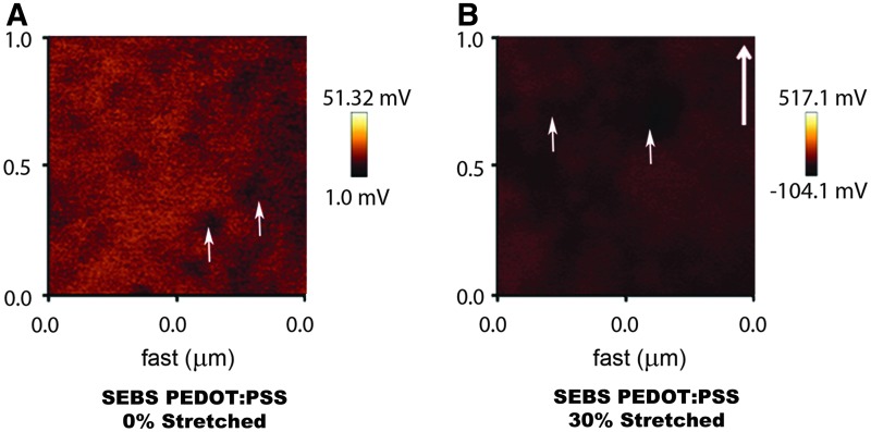
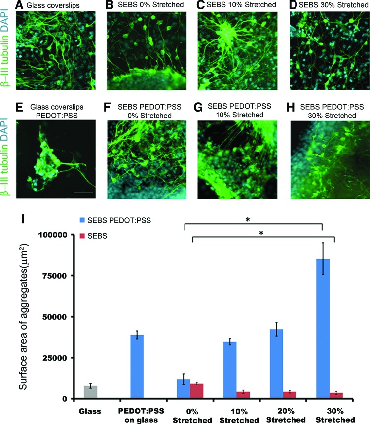
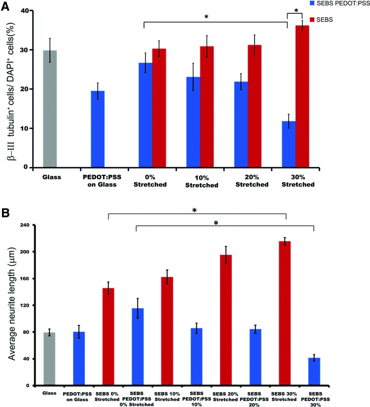
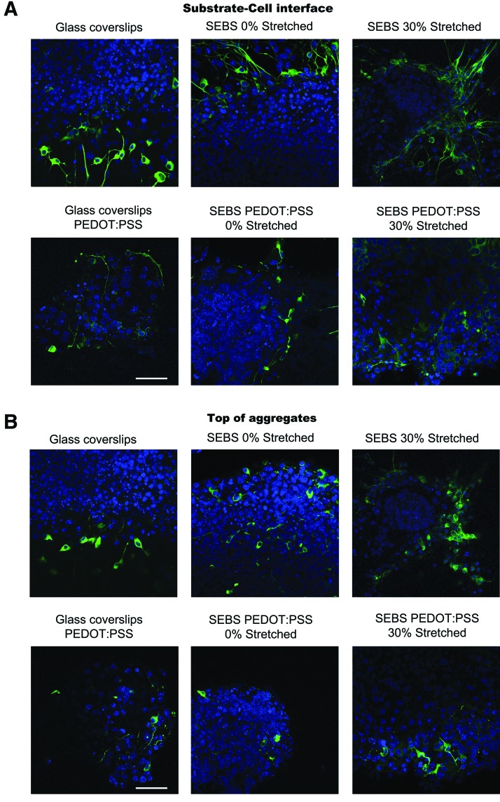
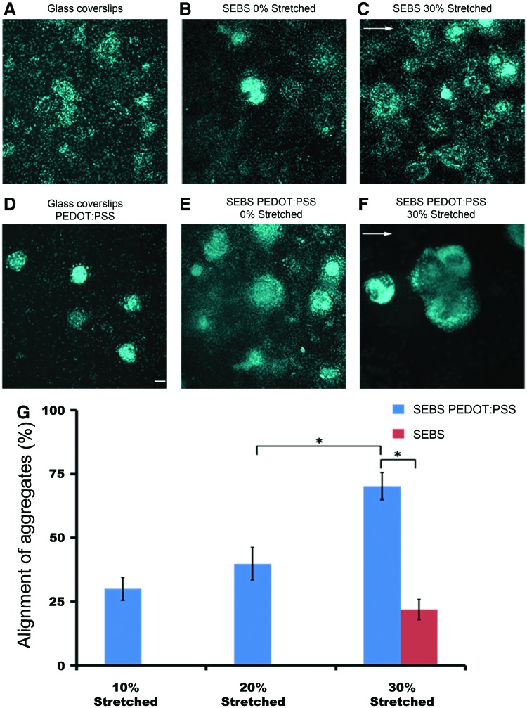
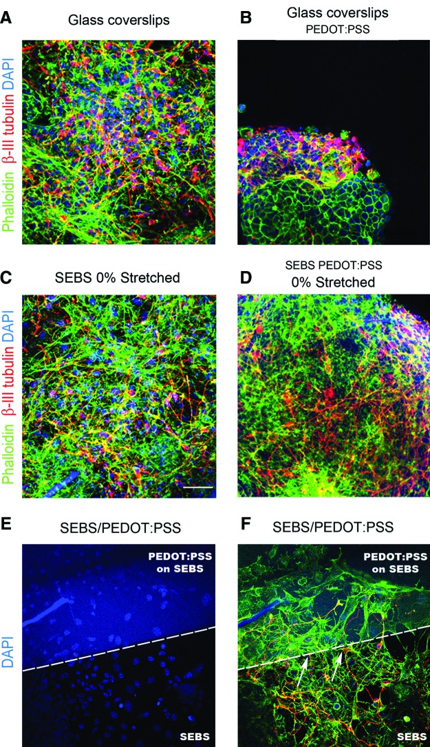
Electrically conducting polymers are prospective candidates as active substrates for the development of neuroprosthetic devices. The utility of these substrates for promoting differentiation of embryonic stem cells paves viable routes for regenerative medicine. Here, we have tuned the electrical and mechanical cues provided to the embryonic stem cells during differentiation by precisely straining the conducting polymer (CP) coated, elastomeric-substrate. Upon straining the substrates, the neural differentiation pattern occurs in form of aggregates, accompanied by a gradient where substrate interface reveals a higher degree of differentiation. The CP domains align under linear stress along with the formation of local defect patterns leading to disruption of actin cytoskeleton of cells, and can provide a mechano-transductive basis for the observed changes in the differentiation. Our results demonstrate that along with biochemical and mechanical cues, conductivity of the polymer plays a major role in cellular differentiation thereby providing another control feature to modulate the differentiation and proliferation of stem cells.


Figures

FIG. 1.
FIG. 1.
Kelvin probe microscopy (KPM) image showing variation in the surface potential of (A) SEBS PEDOT:PSS 0% Stretched (B) SEBS PEDOT:PSS 30% Stretched substrates. Bold arrow indicates the direction of strain, while small arrows indicate the PEDOT domains. The alignment and distribution of PEDOT domains change on straining the substrates. SEBS, styrene ethylene butylene styrene; PEDOT:PSS, (poly(3,4-ethylenedioxythiophene) poly(styrene sulfonate)). Color images available online at www.liebertpub.com/tea
FIG. 2.
FIG. 2.
Immunocytochemistry for differentiation of ES-NPs into neurons (β-III tubulin) on (B–D) SEBS, (F–H) PEDOT:PSS coated SEBS substrates and (A, E) glass-coverslips and PEDOT:PSS coated glass-coverslips (control). (I) Distribution of the surface area of the cell aggregates (nuclear) formed on each substrate (*p<0.05, One-way analysis of variance (ANOVA)). Data are represented as mean±SD, n=3. (A–H): Scale bar 50 μm. Color images available online at www.liebertpub.com/tea
FIG. 3.
FIG. 3.
Substrate dependence of neuronal differentiation and average neurite length. Neurite length was measured by Image J software and only those neurons were considered for sampling which were spread out on the substrates. (A) Distribution of the percentage of β-III tubulin positive cells on various substrates (*p<0.05, One-way ANOVA). (B) Distribution of the average neurite length of the neurons on various substrates (*p<0.05, One-way ANOVA). Data are presented as mean±SD, (n=3). Color images available online at www.liebertpub.com/tea
FIG. 4.
FIG. 4.
Confocal image of cell aggregate density with focal plane at (A) z=0 corresponding to the substrate-cell interface (B) Top of the cell aggregate. Neuronal differentiation occurs at all the strata of the aggregates with extensive neurite branching on strained CP substrates. Scale bar, 50 μm. Color images available online at www.liebertpub.com/tea
FIG. 5.
FIG. 5.
Directional alignment of the cell aggregates along the local defects generated orthogonal to the strain direction (A–F). Arrows in the figure indicate the strain direction. (G) Distribution of the directional alignment of cellular aggregates along the “defect patterns” generated on polymeric substrates (*p<0.05, One-way analysis of variance). Data are represented as mean±SD, (n=3). (A–F): Scale bar, 50 μm. Color images available online at www.liebertpub.com/tea
FIG. 6.
FIG. 6.
Actin cytoskeleton (Phalloidin) of the differentiated cells on polymeric substrates (A–D, F). Regular arrangement of actin cytoskeleton is seen on (A) glass-coverslips (C) SEBS 0% stretched. Disruption of actin fibers occur on PEDOT:PSS coated SEBS substrates leading to rounded morphology and aggregation of cells (B, D). Nucleus (DAPI) of the differentiated cells on the substrate containing SEBS and PEDOT:PSS coated SEBS (E). The arrows in (F) show the neurite ending on the interface of PEDOT: PSS and SEBS. Disruption of actin cytoskeleton is also seen on the conducting side of the patterned substrate. Scale bar, 50 μm. Color images available online at www.liebertpub.com/tea

Comment in

References

    1. Meredith J.E. Fazeli B. Schwartz M.A. The extracellular-matrix as a cell-survival factor. Mol Biol Cell. 1993;4:953. - PMC - PubMed
    1. Colognato H. Winkelmann D.A. Yurchenco P.D. Laminin polymerization induces a receptor-cytoskeleton network. J Cell Biol. 1999;145:619. - PMC - PubMed
    1. Gumbiner B.M. Cell adhesion: the molecular basis of tissue architecture and morphogenesis. Cell. 1996;84:345. - PubMed
    1. Burridge K. Fath K. Kelly T. Nuckolls G. Turner C. Focal adhesions: transmembrane junctions between the extracellular matrix and the cytoskeleton. Annu Rev Cell Biol. 1988;4:487. - PubMed
    1. Dembo M. Wang Y.L. Stresses at the cell-to-substrate interface during locomotion of fibroblasts. Biophys J. 1999;76:2307. - PMC - PubMed