. 2013 May 31:31:38-47.
doi: 10.1016/j.niox.2013.03.006. Epub 2013 Mar 30.

Nitroxyl accelerates the oxidation of oxyhemoglobin by nitrite

Affiliations

Nitroxyl accelerates the oxidation of oxyhemoglobin by nitrite

Landon Bellavia et al. Nitric Oxide. .

Abstract

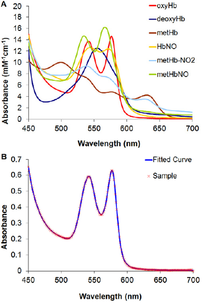
Angeli's salt (Na₂N₂O₃) decomposes into nitroxyl (HNO) and nitrite (NO₂(-)), compounds of physiological and therapeutic interest for their impact on biological signaling both through nitric oxide and nitric oxide independent pathways. Both nitrite and HNO oxidize oxygenated hemoglobin to methemoglobin. Earlier work has shown that HNO catalyzes the reduction of nitrite by deoxygenated hemoglobin. In this work, we have shown that HNO accelerates the oxidation of oxygenated hemoglobin by NO₂(-). We have demonstrated this HNO mediated acceleration of the nitrite/oxygenated hemoglobin reaction with oxygenated hemoglobin being in excess to HNO and nitrite (as would be found under physiological conditions) by monitoring the formation of methemoglobin in the presence of Angeli's salt with and without added NO₂(-). In addition, this acceleration has been demonstrated using the HNO donor 4-nitrosotetrahydro-2H-pyran-4-yl pivalate, a water-soluble acyloxy nitroso compound that does not release NO₂(-) but generates HNO in the presence of esterase. This HNO donor was used both with and without NO₂(-) and acceleration of the NO₂(-) induced formation of methemoglobin was observed. We found that the acceleration was not substantially affected by catalase, superoxide dismutase, c-PTIO, or IHP, suggesting that it is not due to formation of extramolecular peroxide, NO₂ or H₂O₂, or to modulation of allosteric properties. In addition, we found that the acceleration is not likely to be related to HNO binding to free reduced hemoglobin, as we found HNO binding to reduced hemoglobin to be much weaker than has previously been proposed. We suggest that the mechanism of the acceleration involves local propagation of autocatalysis in the nitrite-oxygenated Hb reaction. This acceleration of the nitrite oxyhemoglobin reaction could affect studies aimed at understanding physiological roles of HNO and perhaps nitrite and use of these agents in therapeutics such as hemolytic anemias, heart failure, and ischemia reperfusion injury.


Figures

Figure 1
Figure 1
Spectral deconvolution of hemoglobin species. (A) Basis spectra used to fit sample spectra for determination of hemoglobin species concentrations. Basis spectra include oxygenated Hb (oxyHb), deoxygenated Hb (deoxyHb), methemoglobin (metHb), nitrite bound metHb (metHb-NO2), ferrous nitrosyl Hb (HbNO), and ferric nitrsoyl Hb (metHbNO). (B) A sample spectrum (red crosses) of 1 mM oxyHb + 50 µM AS + 50 µM NO2, incubated for 10 min at 37°C, fit by a least-squares regression to a linear combination of basis spectra (blue line).
Figure 2
Figure 2
The reaction of oxyhemoglobin with Angeli’s salt and/or nitrite. (A) Time-resolved spectra of 1 mM oxyHb upon addition of 50 µM AS and 50 µM NO2 under aerobic conditions. The decrease in absorbance at 542 nm and 577 nm, and the increase in absorbance at 500 nm and 630 nm, demonstrate the partial conversion of oxyHb to metHb. The arrows indicate the direction of spectral shift over time. (B) Methemoglobin levels due to the oxidation of oxyHb by AS and/or nitrite over time. Samples were made with 1 mM oxyHb + 50 µM AS + 50 µM NO2, 1 mM oxyHb + 50 µM AS, 1 mM oxyHb + 50 µM NO2, and 1 mM oxyHb + a blank (consisting of oxyHb alone plus an equivalent amount of NaOH that was added to other samples containing dissolved AS). Spectra were taken every 10 min for an hour, then every hour for four hours. Sample spectra were fit to basis spectra, and the evolution of metHb was tracked. The hemoglobin balance remained oxyHb, with occasional traces (<2.5%) of deoxyHb. No other species were present in quantities detectible by absorption spectroscopy. (C) MetHb formation in 1 mM oxyHb as a result of 50 µM nitrite in the presence (orange) or absence (teal) of 50 µM AS. The metHb concentration in 1 mM oxyHb + 50 µM AS was subtracted from the metHb concentration in 1 mM Hb + 50 µM AS + 50 µM NO2 to yield the metHb concentration due to nitrite in the presence of AS. The metHb concentration in 1 mM oxyHb + 50 µM NO2 was subtracted from the metHb concentration in 1 mM oxyHb + a blank to yield the metHb concentration due to nitrite in the absence of AS. (D) Nitrite levels after 10 min in samples of 1 mM oxyHb + 50 µM AS + 50 µM NO2, 1 mM oxyHb + 50 µM AS, and 1 mM oxyHb + 50 µM NO2, color coded to match the corresponding lines in (B). The rightmost bar shows the nitrite concentration in 1 mM oxyHb + 50 µM AS subtracted from the nitrite concentration in 1 mM oxyHb + 50 µM AS + 50 µM NO2, corresponding to the orange line in (C).
Figure 3
Figure 3
Hemoglobin oxidation by nitrite at different nitrite concentrations. (A) The oxidation of oxyHb to metHb due to 100 µM NO2, 50 µM NO2, and autoxidation. (B) The difference between oxidation due to 100 µM NO2 and 50 µM NO2 compared to the difference between oxidation due to 50 µM NO2 and autoxidation. (C) Methemoglobin levels due to the oxidation of oxyHb by 50 µM DEA NONOate (NO donor) and/or nitrite over time. Conditions are otherwise as in Figure 2B. (D) MetHb formation in 1 mM oxyHb as a result of 50 µM nitrite in the presence (orange) or absence (teal) of 50 µM DEA NONOate. Conditions are otherwise as in Figure 2C.
Figure 4
Figure 4
The reaction of oxyhemoglobin with nitrite and/or HNO from 4-nitrosotetrahydro-2H-pyran-4-yl pivalate, a water-soluble acyloxy nitroso compound that releases HNO in the presence of pig liver esterase. (A) Time-resolved spectra of 1 mM oxyHb upon addition of 50 µM HNO donor and 50 µM NO2 under aerobic conditions. The decrease in absorbance at 542 nm and 577 nm, and the increase in absorbance at 500 nm and 630 nm, demonstrate the partial conversion of oxyHb to metHb. The arrows indicate the direction of spectral shift over time. (B) Methemoglobin levels due to the oxidation of oxyHb by HNO and/or nitrite over time. Samples were made with 1 mM oxyHb + 50 µM HNO donor + 50 µM NO2, 1 mM oxyHb + 50 µM HNO donor, 1 mM oxyHb + 50 µM NO2, and 1 mM oxyHb. Spectra were taken every 10 min for two hours, then once an hour for two more hours. Sample spectra were fit to basis spectra, and the evolution of metHb was monitored. The hemoglobin balance remained oxyHb. No other species were present in quantities detectible by absorption spectroscopy. (C) MetHb formation in 1 mM oxyHb as a result of 50 µM nitrite in the presence (orange) or absence (teal) of 50 µM HNO donor. The metHb concentration in 1 mM oxyHb + 50 µM HNO donor was subtracted from the metHb concentration in 1 mM Hb + 50 µM HNO donor + 50 µM NO2 to yield the metHb concentration due to nitrite in the presence of the HNO donor. The metHb concentration in 1 mM oxyHb + 50 µM NO2 was subtracted from the metHb concentration in 1 mM oxyHb to yield the metHb concentration due to nitrite in the absence of the HNO donor.
Figure 5
Figure 5
The reaction of oxyhemoglobin with Angeli’s salt and/or nitrite in the presence of catalase or c-PTIO. (A) Methemoglobin levels due to the oxidation of oxyHb by AS and/or nitrite over time in the presence of 50 µM catalase. Conditions are otherwise as in Figure 2B. (B) MetHb formation in 1 mM oxyHb with 50 µM catalase as a result of 50 µM nitrite in the presence (orange) or absence (teal) of 50 µM AS. Conditions are otherwise as in Figure 2C. (C) Methemoglobin levels due to the oxidation of oxyHb by AS and/or nitrite over time in the presence of 250 µM c-PTIO. Conditions are otherwise as in Figure 2B. (D) MetHb formation in 1 mM oxyHb with 250 µM c-PTIO as a result of 50 µM nitrite in the presence (orange) or absence (teal) of 50 µM AS. Conditions are otherwise as in Figure 2C.
Figure 6
Figure 6
The reaction of oxyhemoglobin with Angeli’s salt and/or nitrite in the presence of superoxide dismutase (SOD) or inositol hexaphosphate (IHP). (A) Methemoglobin levels due to the oxidation of oxyHb by AS and/or nitrite over time in the presence of 10 Ku/mL SOD. Conditions are otherwise as in Figure 2B. (B) MetHb formation in 1 mM oxyHb with 10 Ku/mL SOD as a result of 50 µM nitrite in the presence (orange) or absence (teal) of 50 µM AS. Conditions are otherwise as in Figure 2C. (C) Methemoglobin levels due to the oxidation of oxyHb by AS and/or nitrite over time in the presence of 2 mM IHP. Conditions are otherwise as in Figure 2B. (D) MetHb formation in 1 mM oxyHb with 2 mM IHP as a result of 50 µM nitrite in the presence (orange) or absence (teal) of 50 µM AS. Conditions are otherwise as in Figure 2C.
Figure 7
Figure 7
The reaction of oxyhemoglobin with Angeli’s salt at differing initial concentrations of metHb and oxyHb. (A) Methemoglobin levels due to the oxidation of oxyHb by AS and/or nitrite over time with an initial concentration of 750 µM oxyHb and 250 µM metHb. Conditions are otherwise as in Figure 2B. (B) MetHb formation in 750 µM oxyHb and 250 µM metHb as a result of 50 µM nitrite in the presence (orange) or absence (teal) of 50 µM AS. Conditions are otherwise as in Figure 2C. (C) Methemoglobin levels due to the oxidation of oxyHb by AS and/or nitrite over time with an initial oxyHb concentration of 200 µM. Conditions are otherwise as in Figure 2B. (D) MetHb formation in 200 µM oxyHb as a result of 50 µM nitrite in the presence (orange) or absence (teal) of 50 µM AS. Conditions are otherwise as in Figure 2C.

References

    1. Andrews KL, Irvine JC, Tare M, Apostolopoulos J, Favaloro JL, Triggle CR, Kemp-Harper BK. A role for nitroxyl (HNO) as an endothelium-derived relaxing and hyperpolarizing factor in resistance arteries. British Journal of Pharmacology. 2009;157:540–550. - PMC - PubMed
    1. Bermejo E, Saenz DA, Alberto F, Rosenstein RE, Bari SE, Lazzari MA. Effect of nitroxyl on human platelets function. Thrombosis and Haemostasis. 2005;94:578–584. - PubMed
    1. Chazotte-Aubert L, Oikawa S, Gilibert I, Bianchini F, Kawanishi S, Ohshima H. Cytotoxicity and site-specific DNA damage induced by nitroxyl anion (NO−) in the presence of hydrogen peroxide - Implications for various pathophysiological conditions. Journal of Biological Chemistry. 1999;274:20909–20915. - PubMed
    1. Cheong E, Tumbev V, Abramson J, Salama G, Stoyanovsky DA. Nitroxyl triggers Ca2+ release from skeletal and cardiac sarcoplasmic reticulum by oxidizing ryanodine receptors. Cell Calcium. 2005;37:87–96. - PubMed
    1. Colton CA, Gbadegesin M, Wink DA, Miranda KM, Espey MG, Vicini S. Nitroxyl anion regulation of the NMDA receptor. Journal of Neurochemistry. 2001;78:1126–1134. - PubMed

Publication types

LinkOut - more resources