Review
. 2013 Apr 1;5(4):a010165.
doi: 10.1101/cshperspect.a010165.

Replication clamps and clamp loaders

Affiliations
Review

Replication clamps and clamp loaders

Mark Hedglin et al. Cold Spring Harb Perspect Biol. .

Abstract

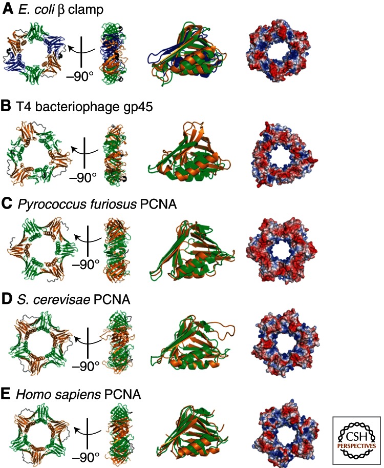
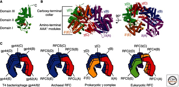
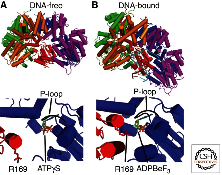
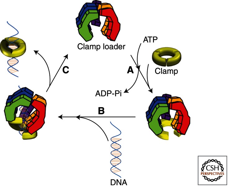
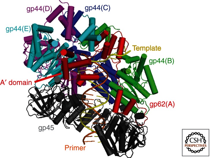
To achieve the high degree of processivity required for DNA replication, DNA polymerases associate with ring-shaped sliding clamps that encircle the template DNA and slide freely along it. The closed circular structure of sliding clamps necessitates an enzyme-catalyzed mechanism, which not only opens them for assembly and closes them around DNA, but specifically targets them to sites where DNA synthesis is initiated and orients them correctly for replication. Such a feat is performed by multisubunit complexes known as clamp loaders, which use ATP to open sliding clamp rings and place them around the 3' end of primer-template (PT) junctions. Here we discuss the structure and composition of sliding clamps and clamp loaders from the three domains of life as well as T4 bacteriophage, and provide our current understanding of the clamp-loading process.


Figures

Figure 1.
Figure 1.
Sliding clamps from different organisms. Crystal structures of (A) the Escherichia coli β clamp (Protein Data Bank [PDB] code 2POL), (B) gene 45 protein (gp45) from T4 bacteriophage (PDB code 1CZD), (C) Pyrococcus furiosus proliferating cell nuclear antigen (PCNA, PDB code 1GE8), (D) Saccharomyces cerevisiae PCNA (PDB code 1PLQ), and (E) Homo sapiens PCNA (PDB code 1AXC). For each organism, the top and side views of the three-dimensional crystal structure are shown on the left and alignment of the globular domains in the middle. The electrostatic surface potential for each is shown on the right with red and blue representing negative and positive electrostatic potentials, respectively. All images were constructed using PyMOL. The sliding clamps from all domains of life have a similar architecture comprised of six domains arranged in a circle. The chain-folding topologies of each domain are the same. The E. coli β clamp is homodimeric (three domains per monomer), whereas T4 gp45 and PCNA from eukaryotes and archaea are each homotrimeric (two domains per monomer). The monomers are arranged head-to-tail in all clamps, resulting in structurally distinct faces. The face from which the carboxyl termini protrude (“carboxyl-terminus face”) interacts with clamp loaders and polymerases. A strong positive electrostatic potential lines the inner surface of each ring where double-stranded DNA resides, favoring nonspecific interactions with the negatively charged phosphate backbone.
Figure 2.
Figure 2.
Clamp loader structure. (A) Close-up of the E. coli γD subunit (from PDB code 1XXH) bound to ATPγS (shown in space-filling format). The carboxyl terminus and the three domains (I, II, and III) are indicated. (B) Front (left) and carboxy-terminal/top (right) views of the γ3δδ′ minimal clamp loader structure (PDB code 1XXH). The “carboxy-terminal collar” and amino-terminal AAA+ ATPase modules are indicated. All images were generated using PyMOL. (C) Stylized images of the clamp loaders from bacteria (γ complex), eukaryotes (RFC), bacteriophage T4 (gp44/62), and archaea (RFC). The positions of the five subunits of each clamp loader complex are denoted by the letters A–E, with analogous subunits indicated by matching colors.
Figure 3.
Figure 3.
DNA promotes ATP hydrolysis by triggering a structural rearrangement of the interfacial ATPase sites of the γ complex. The DNA-free (A, PDB code 1XXH) and DNA-bound (B, PDB code 3GLF) forms of the γ complex are shown. For each, the structure of the γ complex is shown on top with a close-up of the γCB interfacial ATPase site shown at the bottom. All images were generated using PyMOL. Nucleotide is bound in the AAA+ module of the γB subunit. The “P-loop” is indicated and shown in black. In the absence of DNA, the “arginine finger” (Arg169) from the γC subunit is too far away to interact with the γ phosphate of ATP in the γB subunit. On binding DNA, the γ complex adopts a right-handed spiral configuration with a uniform rise and rotation that closely mimics that of the bound DNA. Such a structural rearrangement presents the “arginine finger” (Arg169) from one subunit (γC) to the γ phosphate of ATP bound in the next subunit (γB), promoting ATP hydrolysis.
Figure 4.
Figure 4.
The clamp-loading mechanism. The catalytic reaction cycle for the loading of sliding clamps onto DNA by clamp loaders is shown as a schematic diagram. (A) Opening of the sliding clamp ring. In the absence of ATP, clamp loaders bind their respective clamps very weakly. On binding ATP, clamp loaders undergo a conformational change, which permits optimal interaction with the carboxy-terminal face of their respective clamp and subsequent opening of the clamp ring. (B) PT junction binding. The open clamp–clamp loader complex together specifically recognizes and binds a PT junction, adopting a “notched screw cap arrangement,” which matches the helical geometry of the DNA duplex and properly aligns the interfacial ATPase sites for hydrolysis. (C) Closure of the clamp ring. On hydrolysis of ATP, clamp loaders revert back to a low-affinity DNA-binding state and eject, leaving the PT DNA positioned within an opened sliding clamp ring. Concurrent or subsequent to ejection, electrostatic interactions between the positively charged inner surface of the sliding clamp ring and the negatively charged DNA drives closure of the open sliding clamp around DNA.
Figure 5.
Figure 5.
The clamp–clamp loader–DNA complex from T4 bacteriophage. Structure of the T4 clamp loader (gp44/62) bound to an open clamp (gp45) and PT DNA (from PDB 3U60) generated using PyMOL. PT DNA, gp44/62 (multicolored), and gp45 (gray) are shown in cartoon form. The A′ domain (red) as well as the primer (orange) and template (yellow) strands of the duplex DNA are indicated.

References

    1. Adelman JL, Chodera JD, Kuo IF, Miller TF III, Barsky D 2010. The mechanical properties of PCNA: Implications for the loading and function of a DNA sliding clamp. Biophys J 98: 3062–3069 - PMC - PubMed
    1. Ahmadian MR, Stege P, Scheffzek K, Wittinghofer A 1997. Confirmation of the arginine-finger hypothesis for the GAP-stimulated GTP-hydrolysis reaction of Ras. Nat Struct Biol 4: 686–689 - PubMed
    1. Allen BL, Uhlmann F, Gaur LK, Mulder BA, Posey KL, Jones LB, Hardin SH 1998. DNA recognition properties of the N-terminal DNA binding domain within the large subunit of replication factor C. Nucleic Acids Res 26: 3877–3882 - PMC - PubMed
    1. Alley SC, Jones AD, Soumillion P, Benkovic SJ 1999a. The carboxyl terminus of the bacteriophage T4 DNA polymerase contacts its sliding clamp at the subunit interface. J Biol Chem 274: 24485–24489 - PubMed
    1. Alley SC, Shier VK, Abel-Santos E, Sexton DJ, Soumillion P, Benkovic SJ 1999b. Sliding clamp of the bacteriophage T4 polymerase has open and closed subunit interfaces in solution. Biochemistry 38: 7696–7709 - PubMed

Publication types

LinkOut - more resources