Review
. 2013 Apr 1;5(4):a012633.
doi: 10.1101/cshperspect.a012633.

Postreplicative mismatch repair

Affiliations
Review

Postreplicative mismatch repair

Josef Jiricny. Cold Spring Harb Perspect Biol. .

Abstract

The mismatch repair (MMR) system detects non-Watson-Crick base pairs and strand misalignments arising during DNA replication and mediates their removal by catalyzing excision of the mispair-containing tract of nascent DNA and its error-free resynthesis. In this way, MMR improves the fidelity of replication by several orders of magnitude. It also addresses mispairs and strand misalignments arising during recombination and prevents synapses between nonidentical DNA sequences. Unsurprisingly, MMR malfunction brings about genomic instability that leads to cancer in mammals. But MMR proteins have recently been implicated also in other processes of DNA metabolism, such as DNA damage signaling, antibody diversification, and repair of interstrand cross-links and oxidative DNA damage, in which their functions remain to be elucidated. This article reviews the progress in our understanding of the mechanism of replication error repair made during the past decade.


Figures
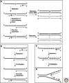
Figure 1.
Figure 1.
General scheme of replication error repair. (A) An error (in this example, G/T mismatch) generated by a DNA polymerase during replication must be repaired before the following round of replication. Unrepaired mismatches are fixed as errors in 50% progeny DNA (top right). MMR-mediated repair involves excision of a tract of the nascent strand (gray) that includes the misincorporated nucleotide and resynthesis of the excised tract. (B) DNA polymerases can generate 12 possible mispairs. Because these cause a misalignment between the primer terminus and template strand, incorporation of the next nucleotide (particularly, the attack of the 3′ OH of the primer terminus on the α phosphate of the incoming deoxyribonucleotide triphosphate [dNTP]) is inefficient. This kinetic barrier allows for translocation of the primer terminus from the polymerase active site to its proofreading exonuclease site, which removes several nucleotides from the primer such that it can realign with the template. Mispairs that escape proofreading are substrates for MMR. (C) Slippage of the primer strand in a repetitive sequence such as a microsatellite will generate an IDL (in this example, a single extrahelical A). If this structure arises behind the polymerase, it will not be detected by the proofreading exonuclease and its repair is thus entirely dependent on MMR. (D) MMR must be directed to the nascent DNA strand that carries—by definition—the erroneous genetic information. In this example, the G/T mismatch must be corrected to G/C.
Figure 2.
Figure 2.
Comparison of overall fold and lesion recognition of E. coli MutS (A, D; 1E3M.pdb), human MutSα (B, E; 2O8B.pdb), and human MutSβ (C, F; 3THY.pdb). (A–C) Ribbon representations of the overall structures. The subunit directly recognizing the DNA mismatch or IDL (subunit A in E. coli MutS, MSH6 in MutSα, or MSH3 in MutSβ) is shown in green; the other subunit (Subunit B, MSH2) is shown in blue. DNA and bound ADP cofactors are depicted in dark red. (D,E) E. coli MutS and human MutSα bind DNA containing a G/T mismatch between their nonspecific clamps and mismatch-binding domains by inducing a 60° kink in the DNA and interacting with the mismatched bases (red) via conserved phenylalanine and glutamate residues (side chains in yellow). (F), Human MutSβ binds DNA containing a two-residue insertion by introducing a 90° kink in the DNA and forming specific interactions with the DNA loop (red) using a lysine and a tyrosine (side chains in yellow). (Figure created using PyMOL [http://www.pymol.org].)
Figure 3.
Figure 3.
Putative conformations of MutSα, viewed down the longitudinal axis of the DNA helix (shown as a cartwheel). (A) On homoduplex DNA, MutSα is loosely bound and follows the winding of the helix. It slowly hydrolyzes ATP, and both subunits can be occupied by either ADP or ATP (shown as A*P). (B) After mismatch detection, the phenylalanine and glutamate of the GxFxE insert into the helix at the mismatch site (shown as rotation of the MSH6 “ratchet” into the DNA). This conformational change triggers an ADP–ATP exchange in MSH6 accompanied by ATPase inhibition. (C) A similar exchange in the MSH2 subunit causes a withdrawal of the ratchet from the DNA. This long-lived clamp is free to slide along the DNA contour.
Figure 4.
Figure 4.
Putative conformations of MutL and its homologs. (A) The proteins are dimerized via their carboxy-terminal domains. (B) DNA binding causes dimerization of the amino-terminal ATPase domains, such that the protein may encircle the helix. For MutL endonucleases, the conformational change accompanying this process may poise the nuclease domain of one of the MutL subunits for cleavage of the sugar-phosphate backbone of the DNA while blocking the nuclease site of the second subunit. (In MutLα, only the PMS subunits harbor nuclease activity.) Cleavage requires activation through interaction with β or PCNA.
Figure 5.
Figure 5.
Schematic representation of the heteroduplex substrates used in in vitro MMR assays. The mismatch (G/T, in this example) is positioned in a restriction enzyme recognition sequence and the plasmid is thus refractory to cleavage by this enzyme. Although covalently closed circular heteroduplexes remain largely refractory to restriction digestion, introduction of a single nick either 5′ (A), or 3′ (B) from the mispaired G results in G/T to A/T correction, as measured by the efficiency of restriction cleavage. In the human system, 5′ → 3′ excision A requires MutSα, EXO1, and RPA. 3′ → 5′ excision B requires, in addition, MutLα, PCNA, and RFC. In the latter system, the mismatch-activated MutSα/MutLα/PCNA complex generates additional nicks in the prenicked strand that are used as EXO1 loading sites for 5′ → 3′ excision of the error-containing strand.
Figure 6.
Figure 6.
MSH6–PCNA interaction. (A) Evolutionary conservation of the eukaryotic PIP motif (bold on grey background). Hs, Homo sapiens; Sc, Saccharomyces cerevisiae; Sp, Schizosaccharomyces pombe; Cg, Cricetulus griseus; Ca, Candida albicans; Mm, Mus musculus; Xt, Xenopus tropicalis; Dr, Danio rerio; At, Arabidopsis thaliana. (B) Schematic representation of human MutSα in complex with PCNA derived from SAXS analysis (Iyer et al. 2008). Although the interaction among these proteins has been believed to be mediated by the PIP motif, PCNA is contacted by many residues of the amino-terminal domain of MSH6, as predicted from experiments with the yeast factor (Shell et al. 2007b). The PIP motif is thus possibly only a docking motif that brings the two polypeptides into close proximity, such that they can interact more intimately. This might help to explain why mutation of PIP motifs of several PCNA interacting proteins often fails to abolish their function.
Figure 7.
Figure 7.
Alternative schemes of MMR in the leading and lagging strands. A mismatch in the leading strand will require the mismatch-activated MutSα/MutLα/PCNA complex to introduce breaks into the nascent strand (left). Once introduced, these breaks can be used as loading sites for EXO1 or, alternatively, as sites of strand displacement by a polymerase loaded at a 3′ terminus upstream of the misincorporated nucleotide (middle). Strand displacement would not require EXO1 but would be dependent on PCNA and the flap-endonuclease FEN1. Mismatch repair in the lagging strand may not absolutely require the MutLα endonuclease, due to the availability of free 5′ termini. MMR in the newest Okazaki fragment will absolutely require EXO1, given that there is no upstream 3′ terminus at which strand displacement might initiate. The repair process might involve the resynthesis of the entire Okazaki fragment (middle).

References

    1. Acharya S, Foster PL, Brooks P, Fishel R 2003. The coordinated functions of the E. coli MutS and MutL proteins in mismatch repair. Mol Cell 12: 233–246 - PubMed
    1. Alani E, Sokolsky T, Studamire B, Miret JJ, Lahue RS 1997. Genetic and biochemical analysis of Msh2p-Msh6p: Role of ATP hydrolysis and Msh2p-Msh6p subunit interactions in mismatch base pair recognition. Mol Cell Biol 17: 2436–2447 - PMC - PubMed
    1. Antony E, Hingorani MM 2003. Mismatch recognition-coupled stabilization of Msh2-Msh6 in an ATP-bound state at the initiation of DNA repair. Biochemistry 42: 7682–7693 - PMC - PubMed
    1. Arana ME, Kunkel TA 2010. Mutator phenotypes due to DNA replication infidelity. Semin Cancer Biol 20: 304–311 - PMC - PubMed
    1. Bai H, Lu A-L 2007. Physical and functional interactions between Escherichia coli MutY glycosylase and mismatch repair protein MutS. J Bacteriol 189: 902–910 - PMC - PubMed

Publication types