

Deer mouse hemoglobin exhibits a lowered oxygen affinity owing to mobility of the E helix
- PMID: 23545644
- PMCID: PMC3614163
- DOI: 10.1107/S1744309113005708
Deer mouse hemoglobin exhibits a lowered oxygen affinity owing to mobility of the E helix
Erratum in
- Acta Crystallogr Sect F Struct Biol Cryst Commun. 2013 Jun;69(Pt 6):710
Abstract
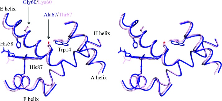
The deer mouse, Peromyscus maniculatus, exhibits altitude-associated variation in hemoglobin oxygen affinity. To examine the structural basis of this functional variation, the structure of the hemoglobin was solved. Recombinant hemoglobin was expressed in Escherichia coli and was purified by ion-exchange chromatography. Recombinant hemoglobin was crystallized by the hanging-drop vapor-diffusion method using polyethylene glycol as a precipitant. The obtained orthorhombic crystal contained two subunits in the asymmetric unit. The refined structure was interpreted as the aquo-met form. Structural comparisons were performed among hemoglobins from deer mouse, house mouse and human. In contrast to human hemoglobin, deer mouse hemoglobin lacks the hydrogen bond between α1Trp14 in the A helix and α1Thr67 in the E helix owing to the Thr67Ala substitution. In addition, deer mouse hemoglobin has a unique hydrogen bond at the α1β1 interface between residues α1Cys34 and β1Ser128.
Keywords: Peromyscus maniculatus; deer mouse; hemoglobin; oxygen affinity.
Figures
Similar articles
-
Alteration of the α1β2/α2β1 subunit interface contributes to the increased hemoglobin-oxygen affinity of high-altitude deer mice.PLoS One. 2017 Mar 31;12(3):e0174921. doi: 10.1371/journal.pone.0174921. eCollection 2017. PLoS One. 2017. PMID: 28362841 Free PMC article.
-
Evolutionary and functional insights into the mechanism underlying high-altitude adaptation of deer mouse hemoglobin.Proc Natl Acad Sci U S A. 2009 Aug 25;106(34):14450-5. doi: 10.1073/pnas.0905224106. Epub 2009 Aug 10. Proc Natl Acad Sci U S A. 2009. PMID: 19667207 Free PMC article.
-
Parakeet Hemoglobin - Its Crystal Structure and Oxygen Affinity in Relation to Some Avian Hemoglobins.Protein Pept Lett. 2021;28(1):18-30. doi: 10.2174/0929866527666200320100109. Protein Pept Lett. 2021. PMID: 32196438
-
Crystal Structure Analysis of Great Cormorant (Phalacrocorax carbo) Hemoglobin to Understand its High Oxygen Affinity Characteristics by Special Structural Features.Protein Pept Lett. 2018;25(8):748-756. doi: 10.2174/0929866525666180620163632. Protein Pept Lett. 2018. PMID: 29929459
-
A novel low oxygen affinity recombinant hemoglobin (alpha96val--> Trp): switching quaternary structure without changing the ligation state.J Mol Biol. 1995 May 12;248(4):867-82. doi: 10.1006/jmbi.1995.0267. J Mol Biol. 1995. PMID: 7752247
Cited by
-
Bohr effect and temperature sensitivity of hemoglobins from highland and lowland deer mice.Comp Biochem Physiol A Mol Integr Physiol. 2016 May;195:10-4. doi: 10.1016/j.cbpa.2016.01.018. Epub 2016 Jan 22. Comp Biochem Physiol A Mol Integr Physiol. 2016. PMID: 26808972 Free PMC article.
-
Epistasis among adaptive mutations in deer mouse hemoglobin.Science. 2013 Jun 14;340(6138):1324-7. doi: 10.1126/science.1236862. Science. 2013. PMID: 23766324 Free PMC article.
-
Crystal structure of hemoglobin from mouse (Mus musculus) compared with those from other small animals and humans.Acta Crystallogr F Struct Biol Commun. 2021 Apr 1;77(Pt 4):113-120. doi: 10.1107/S2053230X2100306X. Epub 2021 Apr 1. Acta Crystallogr F Struct Biol Commun. 2021. PMID: 33830076 Free PMC article.
-
Alteration of the α1β2/α2β1 subunit interface contributes to the increased hemoglobin-oxygen affinity of high-altitude deer mice.PLoS One. 2017 Mar 31;12(3):e0174921. doi: 10.1371/journal.pone.0174921. eCollection 2017. PLoS One. 2017. PMID: 28362841 Free PMC article.
-
Intraspecific polymorphism, interspecific divergence, and the origins of function-altering mutations in deer mouse hemoglobin.Mol Biol Evol. 2015 Apr;32(4):978-97. doi: 10.1093/molbev/msu403. Epub 2015 Jan 2. Mol Biol Evol. 2015. PMID: 25556236 Free PMC article.
References
-
- Adams, P. D. et al. (2010). Acta Cryst. D66, 213–221. - PubMed
-
- Antonini, E. & Brunori, M. (1971). Hemoglobin and Myoglobin in their Reactions with Ligands Amsterdam: North-Holland.
Publication types
MeSH terms
Substances
Associated data
- Actions
Grants and funding
LinkOut - more resources
Full Text Sources
Other Literature Sources
Miscellaneous
