. 2013 Apr 1;69(Pt 4):399-404.
doi: 10.1107/S1744309113005939. Epub 2013 Mar 29.

Structure of an atypical FeoB G-domain reveals a putative domain-swapped dimer

Affiliations

Structure of an atypical FeoB G-domain reveals a putative domain-swapped dimer

Chandrika N Deshpande et al. Acta Crystallogr Sect F Struct Biol Cryst Commun. .

Abstract

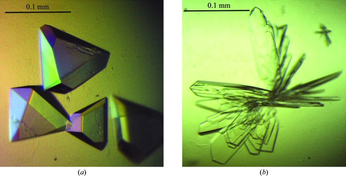
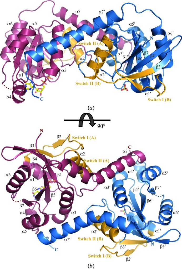
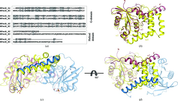
FeoB is a transmembrane protein involved in ferrous iron uptake in prokaryotic organisms. FeoB comprises a cytoplasmic soluble domain termed NFeoB and a C-terminal polytopic transmembrane domain. Recent structures of NFeoB have revealed two structural subdomains: a canonical GTPase domain and a five-helix helical domain. The GTPase domain hydrolyses GTP to GDP through a well characterized mechanism, a process which is required for Fe(2+) transport. In contrast, the precise role of the helical domain has not yet been fully determined. Here, the structure of the cytoplasmic domain of FeoB from Gallionella capsiferriformans is reported. Unlike recent structures of NFeoB, the G. capsiferriformans NFeoB structure is highly unusual in that it does not contain a helical domain. The crystal structures of both apo and GDP-bound protein forms a domain-swapped dimer.

Keywords: FeoB; Gallionella capsiferriformans; NFeoB; domain-swapped dimer.


Figures

Figure 1
Figure 1
Crystals of G. capsiferriformans NFeoB. (a) Crystals of the apo form grown using condition H2 of The JCSG+ Suite [1 M ammonium sulfate, 0.1 M bis-tris pH 5.5, 1%(w/v) PEG 3350]. (b) Crystals of the GDP-bound form obtained using condition H7 of The JCSG+ Suite [0.2 M ammonium sulfate, 0.1 M bis-tris pH 5.5, 25%(w/v) PEG 3350].
Figure 2
Figure 2
Overall structure of the G. capsiferriformans GDP-bound NFeoB dimer. (a) Ribbon representation of the GDP-bound dimer viewed from the side. Individual protomers are coloured purple (chain A) and blue (chain B). The switch regions are shaded in orange. Loop regions of poor density connecting β7 to α6 (residues 144–147) are represented by dotted lines. A bound molecule of GDP and a sulfate ion occupying the nucleotide-binding site in chain A and chain B, respectively, are represented by sticks and coloured by atom type (C, yellow; O, red; N, blue; P, yellow). (b) Rotated view of (a).
Figure 3
Figure 3
A 2F oF c map around the GDP molecule (ball-and-stick representation coloured by atom type as in Fig. 2 ▶) contoured at 1σ. The protein backbone is shown as grey ribbons. The electron density is shown in light blue. The residues forming hydrogen-bonding interactions with the GDP molecule are shown in stick representation (grey) and are coloured red for oxygen and blue for nitrogen. Hydrogen bonds are shown as black dashed lines, and green spheres represent the waters in the GDP-binding network.
Figure 4
Figure 4
Amino-acid sequence alignment and structural comparison of G. capsiferriformans NFeoB and E. coli NFeoB. (a) Sequence alignment of E. coli NFeoB and G. capsiferriformans NFeoB, with the conserved residues shaded in grey. (b) Structural superimposition of G. capsiferriformans NFeoB chain A (coloured as in Fig. 2 ▶; PDB entry 3w5j) with E. coli NFeoB (yellow; PDB entry 3hyt), illustrating the absence of a large portion of the helical domain in the former. (c) and (d) show structural superimpositions of the G. capsiferriformans NFeoB dimer (views and colours as in Fig. 2 ▶) with E. coli NFeoB, illustrating the analogous positioning of the C-terminal extended helix in the two structures.

References

    1. Afonine, P. V., Grosse-Kunstleve, R. W. & Adams, P. D. (2005). CCP4 Newsl. Protein Crystallogr. 42, contribution 8.
    1. Ali, M. H. & Imperiali, B. (2005). Bioorg. Med. Chem. 13, 5013–5020. - PubMed
    1. Ash, M. R., Guilfoyle, A., Clarke, R. J., Guss, J. M., Maher, M. J. & Jormakka, M. (2010). J. Biol. Chem. 285, 14594–14602. - PMC - PubMed
    1. Ash, M.-R., Maher, M. J., Guss, J. M. & Jormakka, M. (2011a). Acta Cryst. D67, 973–980. - PubMed
    1. Ash, M.-R., Maher, M. J., Guss, J. M. & Jormakka, M. (2011b). Acta Cryst. F67, 1511–1515. - PMC - PubMed

Publication types