Review
. 2013 Apr;143(4):1151-1162.
doi: 10.1378/chest.12-1215.

Balancing the risks and benefits of oxygen therapy in critically III adults

Affiliations
Review

Balancing the risks and benefits of oxygen therapy in critically III adults

G R Scott Budinger et al. Chest. 2013 Apr.

Abstract

Oxygen therapy is an integral part of the treatment of critically ill patients. Maintenance of adequate oxygen delivery to vital organs often requires the administration of supplemental oxygen, sometimes at high concentrations. Although oxygen therapy is lifesaving, it may be associated with deleterious effects when administered for prolonged periods at high concentrations. Here, we review the recent advances in our understanding of the molecular responses to hypoxia and high levels of oxygen and review the current guidelines for oxygen therapy in critically ill patients.


Figures

Figure 1.
Figure 1.
Stabilization of HIF during hypoxia. During normoxia, HIF-1α is hydroxylated by cellular PHDs at specific proline residues. The hydroxylated HIF-1α molecule is recognized by its ubiquitin ligase, the VHL, which targets it for degradation in the proteasome. A fall in the local tissue oxygen concentration inhibits the activity of the PHDs, allowing for the stabilization of HIF-1α, which forms a complex with HIF-1β (also known as ARNT), which is translocated to the nucleus, where it binds to HREs in the genome to regulate the expression of hundreds of genes, including VEGF. ARNT = aryl-hydrocarbon nuclear translocator; HIF = hypoxia inducible factor; HRE = hypoxia inducible factor responsive element; OH = hydroxyl; PHD = prolyl hydroxylase; Ub = ubiquitin; VEGF = vascular endothelial growth factor; VHL = von Hippel Landau protein.
Figure 2.
Figure 2.
Mechanisms of hyperoxia-induced lung injury. Exposure to hyperoxia results in the generation of O2 from the mitochondria and from the NAD(P)H oxidase complex. O2 can be converted to H2O2 and water in a reaction catalyzed by SOD1, which is localized to the mitochondrial intermembrane space and cytosol, or SOD2, which is localized to the mitochondrial matrix. H2O2 is further reduced to oxygen and water by the GPx and by catalase expressed in the cytosol and mitochondria (indicated by *). H2O2 can activate adaptive signaling pathways in the cell, but when present in excess, both O2 and H2O2 can oxidize cellular proteins and lipids or generate ONOO and ˙OH to cause cellular injury. GPx = glutathione peroxidases; H2O2 = hydrogen peroxide; NO = nitric oxide; O2 = superoxide anions; ˙OH = hydroxyl radicals; ONOO = reactive nitrogen species; Prx(s) = peroxiredoxins; SOD = superoxide dismutase.
Figure 3.
Figure 3.
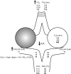
Correction of arterial hypoxemia in patients with alveolar flooding (shunt). The lungs of a patient with an alveolar flooding process (eg, ARDS, cardiogenic pulmonary edema, and so forth) are represented by two alveoli. When the patient is breathing room air (FIO2 =0.21, black numbers), the CvO2 is determined primarily by the amount of oxygen carried on hemoglobin (about 15 mL O2/dL) with a nearly negligible contribution from dissolved oxygen (about 0.15 mL O2/dL). Hemoglobin in the blood flowing through the normal alveoli (unshaded) is fully saturated (about 20 mL O2/dL on Hg and 0.3 mL O2/dL dissolved); however, the oxygen content of the mixed venous blood flowing through the fluid-filled alveoli (shaded) is not changed. With a Q˙S/Q˙T of 0.5 as shown, the resulting arterial oxygen content in room air is 17.5 mL O2/dL, corresponding to an SaO2 of about 89%. Supplemental oxygen therapy can only improve SaO2 by increasing the levels of oxygen dissolved in the plasma. Because these levels are negligible until FIO2 levels approach 1.0 (an FIO2 of 0.3 is shown as an example, italic numbers), shunt physiology is often recognized clinically by the failure to respond to low-flow oxygen or by a reduction in the PaO2/FIO2 ratio. SaO2 in these patients can be improved (black arrows) by increasing the FIO2 to values near 1 (bold numbers), reducing the shunt or increasing the CvO2. CvO2 (mL O2/dL) = %sat × [Hg](g) × (1.34 mL O2/g Hg) + PO2 (torr) × solubility of oxygen in plasma (0.003 mL O2/torr). CvO2 = mixed venous oxygen content; Q˙S/Q˙T = shunt fraction; SaO2 = arterial oxygen saturation. (Adapted with permission from Leff and Schumacker.)
Figure 4.
Figure 4.
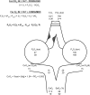
Correction of arterial hypoxemia in patients with alveolar hypoventilation. The difference between the inspired PO2 and the alveolar PO2 is defined by the alveolar gas equation, which is a statement of the conservation of mass for CO2 and oxygen around the alveolus in steady state. Alveolar hypoventilation can result from a fall in minute ventilation or ventilation-perfusion mismatching with increased dead space ventilation. As a result, the alveolar PO2 can approach mixed venous values (an obstructed alveolus with a PO2 similar to the mixed venous is shown). Low-level oxygen therapy results in small increases in the alveolar PO2; however, because hemoglobin binds avidly to oxygen in this range of PO2, these changes are usually sufficient to correct the arterial hypoxemia (an FIO2 of 0.3 is shown in italic numbers). CaO2 = arterial oxygen content; Patm = atmospheric pressure; PH2O = water vapor pressure. See Figure 3 legend for expansion of other abbreviations. (Adapted with permission from Leff and Schumacker.)

References

    1. Priestley J. The Discovery of Oxygen, Part 1. Experiments by Joseph Priestly, LL.D. (1775). Chicago, IL: Albembic Club Reprints: University of Chicago Press; 1912:53-54
    1. Bean JW. Effects of oxygen at increased pressure. Physiol Rev. 1945;25(1):1-147
    1. Becker-Freyseng H, Clamann HG. On the question of oxygen poisoning. J Mol Med. 1939;18(43):1382-1385
    1. Caldwell PR, Lee WL, Jr, Schildkraut HS, Archibald ER. Changes in lung volume, diffusing capacity, and blood gases in men breathing oxygen. J Appl Physiol. 1966;21(5):1477-1483 - PubMed
    1. O’Driscoll BR, Howard LS, Davison AG; British Thoracic Society BTS guideline for emergency oxygen use in adult patients. Thorax. 2008;63(suppl 6):vi1-vi68 - PubMed

Publication types