. 2013 Apr 8;210(4):821-38.
doi: 10.1084/jem.20122005. Epub 2013 Apr 1.

Resetting translational homeostasis restores myelination in Charcot-Marie-Tooth disease type 1B mice

Affiliations

Resetting translational homeostasis restores myelination in Charcot-Marie-Tooth disease type 1B mice

Maurizio D'Antonio et al. J Exp Med. .

Abstract

P0 glycoprotein is an abundant product of terminal differentiation in myelinating Schwann cells. The mutant P0S63del causes Charcot-Marie-Tooth 1B neuropathy in humans, and a very similar demyelinating neuropathy in transgenic mice. P0S63del is retained in the endoplasmic reticulum of Schwann cells, where it promotes unfolded protein stress and elicits an unfolded protein response (UPR) associated with translational attenuation. Ablation of Chop, a UPR mediator, from S63del mice completely rescues their motor deficit and reduces active demyelination by half. Here, we show that Gadd34 is a detrimental effector of CHOP that reactivates translation too aggressively in myelinating Schwann cells. Genetic or pharmacological limitation of Gadd34 function moderates translational reactivation, improves myelination in S63del nerves, and reduces accumulation of P0S63del in the ER. Resetting translational homeostasis may provide a therapeutic strategy in tissues impaired by misfolded proteins that are synthesized during terminal differentiation.


Figures
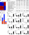
Figure 1.
Figure 1.
Transcriptional profiling reveals a robust stress response in S63del nerves. (A) Hierarchical clustering of P5 sciatic nerves from WT, Chop-null, and P0S63del mice. (B) The number of genes that vary in S63del at different stages of myelination. (C) L2L analysis of the S63del group of genes up-regulated in P5 nerves shows an enrichment for the GO categories related to stress response. (D) Quantitative RT-PCR analysis on P28 sciatic nerves to validate a selection of UPR/ERAD-related genes, and early Schwann cell transcription factors. Error bars, SEM; n = 4–6 RT from independent pools of P28 nerves. *, P < 0.05; **, P < 0.01, Student’s t test.
Figure 2.
Figure 2.
The lipid/cholesterol biosynthetic program is strongly down-regulated in S63del nerves. (A) L2L analysis at P5 of genes down-regulated in S63del nerves identified an enrichment for GO categories related to lipid metabolism. (B) Quantitative RT-PCR for a selection of enzymes involved in the cholesterol biosynthetic pathway. Error bars, SEM; n = 4 RT from independent pools of nerves. *, P < 0.05, **, P < 0.01 by Student’s t test; n.s. = not significant.
Figure 3.
Figure 3.
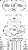
Gadd34 is a target of CHOP in S63del nerves. (A) A Venn diagram of genes up-regulated both in sciatic nerves from P28 WT mice injected with tunicamycin and from P28 S63del mice identified 103 genes in common. Of these, nine returned to WT levels in Chop-null mice injected with tunicamycin and in S63del/Chop-null nerves. (B) Expression of CHOP and the eight CHOP target genes. Exp. Levels = raw level of expression in S63del nerves.
Figure 4.
Figure 4.
Reduction of Gadd34 levels results in increased eIF2α phosphorylation. (A) Quantitative RT-PCR for Gadd34 mRNA from P28 sciatic nerves. Error bars, SEM; *, P < 0.05; n = 4 RT from independent pools of nerves. (B) Western blot analysis on P28 sciatic nerve lysates was performed for Gadd34 and phosphorylated eIF2α; β-tubulin provides a control for loading. One representative experiment of four is shown. Numbers indicate relative molecular weights. (C) Gadd34 and (D) phosphorylated eIF2α protein levels as determined by densitometry. Error bars, SEM; **, P < 0.01, ***, P < 0.001; n = 4.
Figure 5.
Figure 5.
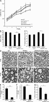
Inactivation of Gadd34 restores motor function and ameliorates the neurophysiological and morphological deficits in S63del mice. (A) Rotarod analysis of motor function in WT, Gadd34Δc/Δc, S63del and S63del/Gadd34Δc/Δc. Error bars, SEM; *, P < 0.05; **, P < 0.01 for S63del/Gadd34Δc/Δc relative to S63del; n = 20–25 animals. (B and C) Electrophysiological analysis showing NCV and F-wave latency; n = 10–12 animals; *, P < 0.05; **, P < 0.01, relative to S63del by Student’s t test. NCV, nerve conduction velocity. (D) Semithin sections stained with toluidine blue from P28 and P180 WT, S63del, and S63del/Gadd34Δc/Δc sciatic nerves. Arrows, amyelinated axons in P28 S63del nerve. Arrowhead, demyelinated fiber/onion bulb in 6-mo-old S63del nerve. Bar, 10 µm. (E) Number of amyelinated axons at P28, and of demyelinated fibers (F) and onion bulbs (G) at 6 mo. Error bars, SEM; *, P < 0.05; **, P < 0.01; ***, P < 0.001, by Student’s t test; n = 20–35 microscopic fields from 4–6 animals per genotype.
Figure 6.
Figure 6.
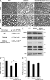
Inactivation of Gadd34 ameliorates the hypomyelination in S63del mice. (A) Semithin sections stained with toluidine blue from P28 and P180 WT, S63del, and S63del/Gadd34Δc/Δc sciatic nerves. Bar, 20 µm. (B) G-ratios in 6-mo old nerves. **, P < 0.01, relative to S63del, by Student’s t test; 8–10 microscopic fields per mouse were analyzed, from 5 mice per genotype. (C) Western blot analysis was performed on sciatic nerve lysates for the myelin proteins MAG, PMP22, and MBP. Numbers indicate relative molecular weights. One representative experiment of three is shown. (D) MBP and (E) PMP22 protein levels as measured by densitometric analysis and E. Error bars, SEM; **, P < 0.01; n = 3 repeat blots.
Figure 7.
Figure 7.
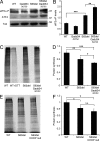
Increased translational attenuation in S63del/Gadd34Δc/Δc nerves. (A) Western blot analysis on P28 sciatic nerves for phosphorylated eIF2α and ATF4. Numbers indicate relative molecular weights. One representative experiment of five is shown. (B) Phosphorylated eIF2α levels, as measured by densitometric analysis. Error bars, SEM; n = 5 repeat blots. (C) Measurement of newly synthesized proteins via S35 incorporation (metabolic labeling) in explanted P28 WT, S63del, and S63del/Gadd34Δc/Δc sciatic nerves. (E) Metabolic labeling measuring newly synthesized proteins in explanted sciatic nerves from P28 WT, Chop-null, and S63del/Chop-null nerves. 1 representative blot of 10 is shown for each experiment. (D and F). Quantification of newly synthesized proteins (via S35 incorporation) by densitometric analysis. (F) Error bars, SEM. *, P < 0.05; **, P < 0.01; ***, P < 0.001, n.s., not significant, by Student’s t test; n = 10 independent experiments.
Figure 8.
Figure 8.
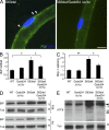
Attenuation of ER stress in S63del/Gadd34Δc/Δc nerves. (A) Immunostaining of teased fibers reveals P0 (green) staining in the perinuclear area (arrowheads) of S63del Schwann cells; nuclei were stained with DAPI (blue). n = 3 mice per genotype were analyzed. Bar, 10 µm. (B and C) mRNA levels for BiP and spliced Xbp-1 were measured by quantitative RT-PCR in P28 sciatic nerves. Error bars, SEM. *, P < 0.05; **, P < 0.01, Student’s t test, n = 6 RT from independent pools of nerves (D and E) Western blot analysis on sciatic nerve lysates for BiP and ATF6. Arrowheads in E indicate the 90-kD precursor and the 50-kD active form of ATF6. Numbers indicate relative molecular weights. One representative experiment of four is shown.
Figure 9.
Figure 9.
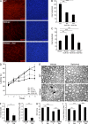
Salubrinal improves myelination in S63del DRG explant cultures and reduces demyelination in S63del mice. (A) Dorsal root ganglia (DRG) were dissected from E14 WT and S63del embryos and myelination was induced with 50 µg/ml ascorbic acid. Myelin internodes were detected with antibodies against MBP (red). Nuclei were visualized with DAPI (blue). Bar, 100 µm. (B and C) Number of myelinating internodes in WT and S63del DRG treated for 3 wk with salubrinal at the indicated concentrations; n = 25–50 fields from 6–8 DRG per condition. Error bars, SEM; **, P < 0.01; ***, P < 0.001. (D) Rotarod analysis of WT and S63del mice treated for 90 d with either salubrinal (Sal) or vehicle (V). Error bars, SEM; n = 12 mice per condition. (E) Semithin sections stained with toluidine blue from P180 sciatic nerves from WT and S63del mice treated with either vehicle (V) or salubrinal (Sal) for 150 d. S63del nerves show several demyelinated fibers (arrowhead) and onion bulbs (arrow). Nerves from 10 mice per genotype were analyzed; bar, 10 µm. (F–G) Percentage of onion bulbs and demyelinated fibers in sciatic nerves from 6-mo-old S63del mice treated with Sal for 150 d. Error bars, SEM; *, P < 0.05; **, P < 0.01 by Student’s t test. 8 microscopic fields from each mouse were analyzed, n = 10 mice per condition. (H and I). Electrophysiological analysis of NCV and F-wave latency in 6-mo-old mice (150 d of treatment with Sal or vehicle). Error bars, SEM; ***, P < 0.001; n.s. = not significant by Student’s t test. n = 12–18 nerves per condition.

Similar articles

Cited by

References

    1. Araki T., Nagarajan R., Milbrandt J. 2001. Identification of genes induced in peripheral nerve after injury. Expression profiling and novel gene discovery. J. Biol. Chem. 276:34131–34141 10.1074/jbc.M104271200 - DOI - PubMed
    1. Arthur-Farraj P.J., Latouche M., Wilton D.K., Quintes S., Chabrol E., Banerjee A., Woodhoo A., Jenkins B., Rahman M., Turmaine M., et al. 2012. c-Jun reprograms Schwann cells of injured nerves to generate a repair cell essential for regeneration. Neuron. 75:633–647 10.1016/j.neuron.2012.06.021 - DOI - PMC - PubMed
    1. Back S.H., Scheuner D., Han J., Song B., Ribick M., Wang J., Gildersleeve R.D., Pennathur S., Kaufman R.J. 2009. Translation attenuation through eIF2alpha phosphorylation prevents oxidative stress and maintains the differentiated state in beta cells. Cell Metab. 10:13–26 10.1016/j.cmet.2009.06.002 - DOI - PMC - PubMed
    1. Barbosa-Tessmann I.P., Chen C., Zhong C., Schuster S.M., Nick H.S., Kilberg M.S. 1999. Activation of the unfolded protein response pathway induces human asparagine synthetase gene expression. J. Biol. Chem. 274:31139–31144 10.1074/jbc.274.44.31139 - DOI - PubMed
    1. Berger P., Niemann A., Suter U. 2006. Schwann cells and the pathogenesis of inherited motor and sensory neuropathies (Charcot-Marie-Tooth disease). Glia. 54:243–257 10.1002/glia.20386 - DOI - PubMed

Publication types