Review
. 2013 Apr;193(4):1025-64.
doi: 10.1534/genetics.112.145219.

DNA repair mechanisms and the bypass of DNA damage in Saccharomyces cerevisiae

Affiliations
Review

DNA repair mechanisms and the bypass of DNA damage in Saccharomyces cerevisiae

Serge Boiteux et al. Genetics. 2013 Apr.

Abstract

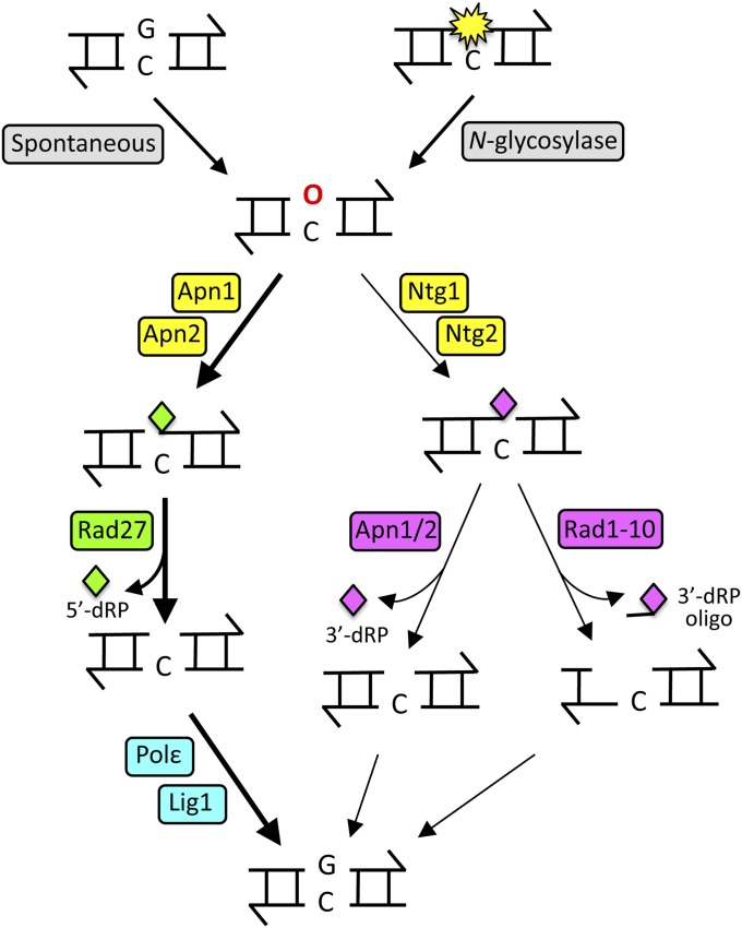
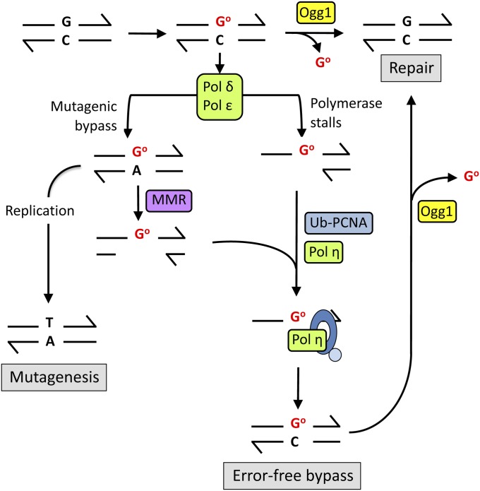
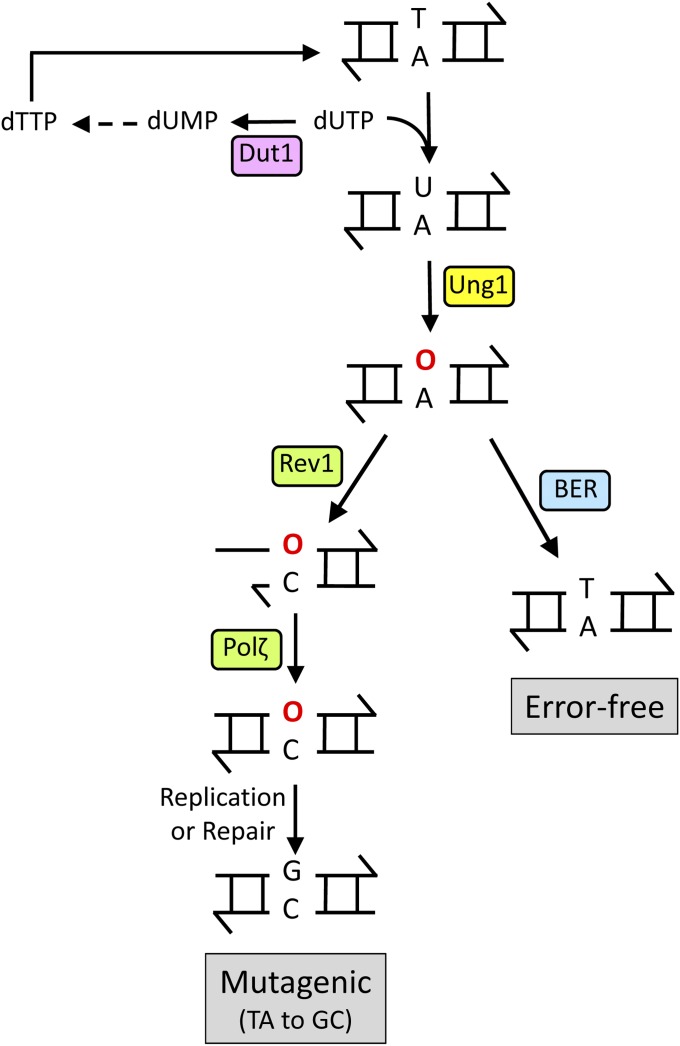
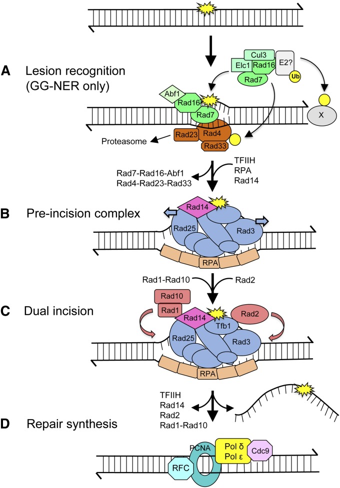
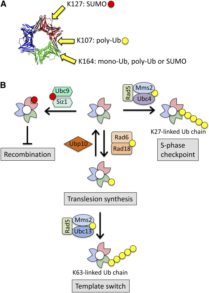
DNA repair mechanisms are critical for maintaining the integrity of genomic DNA, and their loss is associated with cancer predisposition syndromes. Studies in Saccharomyces cerevisiae have played a central role in elucidating the highly conserved mechanisms that promote eukaryotic genome stability. This review will focus on repair mechanisms that involve excision of a single strand from duplex DNA with the intact, complementary strand serving as a template to fill the resulting gap. These mechanisms are of two general types: those that remove damage from DNA and those that repair errors made during DNA synthesis. The major DNA-damage repair pathways are base excision repair and nucleotide excision repair, which, in the most simple terms, are distinguished by the extent of single-strand DNA removed together with the lesion. Mistakes made by DNA polymerases are corrected by the mismatch repair pathway, which also corrects mismatches generated when single strands of non-identical duplexes are exchanged during homologous recombination. In addition to the true repair pathways, the postreplication repair pathway allows lesions or structural aberrations that block replicative DNA polymerases to be tolerated. There are two bypass mechanisms: an error-free mechanism that involves a switch to an undamaged template for synthesis past the lesion and an error-prone mechanism that utilizes specialized translesion synthesis DNA polymerases to directly synthesize DNA across the lesion. A high level of functional redundancy exists among the pathways that deal with lesions, which minimizes the detrimental effects of endogenous and exogenous DNA damage.


Figures

Figure 1
Figure 1
The BER pathway. AP sites (red “O”) are generated either by spontaneous base loss or a DNA N-glycosylase. Apn1 and Apn2 nick the backbone on the 5′ side of an AP site to initiate the major pathway for repair; the resulting 5′-dRP is removed by the Rad27 5′-flap endonuclease. AP-site processing can also be initiated by the Ntg1 or Ntg2 lyase, which nicks on the 3′ side of lesion. The resulting 3′-dRP can be removed by the 3′-diesterase activity of Apn1/Apn2 or as part of a Rad1-Rad10 generated oligonucleotide. Finally, the gap is filled by DNA Pol ε, and the backbone is sealed by DNA ligase 1.
Figure 2
Figure 2
The GO network. Reactive oxygen species attack guanine base-paired with cytosine to yield 8-oxoG (GO). Ogg1 excises 8-oxoG from the DNA backbone, and the resulting AP site is repaired via BER (“repair”). If encountered during replication, local sequence context will determine whether Pol δ/ε stalls at or bypasses the GO lesion. If Pol δ/ε stalls at 8-oxoG during replication, Pol η is recruited by ubiquitinated PCNA (Ub-PCNA) and preferentially incorporates C opposite the lesion (“error-free bypass”). During bypass by Pol δ/ε, adenine is frequently inserted instead of cytosine to create a GO:A mispair, which is recognized by the MMR machinery. The newly synthesized, A-containing strand is degraded to generate a single-strand gap containing the lesion, and C is incorporated opposite the lesion during a gap-filling reaction, which may involve Pol η. If not repaired, the GO:A mispair will yield a GC-to-TA transversion at the next round of replication (“mutagenesis”).
Figure 3
Figure 3
Bypass of endogenous AP sites. dUTP levels in the nucleotide pool are reduced by Dut1 activity, thereby limiting the incorporation of dUMP into genomic DNA. Most endogenous AP sites (indicated by red “O”) are generated by Ung1 removal of uracil in DNA. The resulting AP site can be repaired by the BER pathway or bypassed by the concerted action of Rev1, which usually inserts cytosine opposite the AP site, and Pol ζ, which extends the O:C terminus.
Figure 4
Figure 4
The NER pathway. (A) During GG-NER, a helix-distorting lesion (yellow star) is recognized by Rad4-Rad23-Rad33 and Rad7-Rad16 complexes. Rad7-Rad16-Abf1 has chromatin-remodeling activity, whereas Rad7-Rad16-Elc1-Cul3-E2 has Ub ligase activity, which modifies Rad4 and additional factors (“X”). These reactions allow efficient recognition of lesions by Rad4, its proper positioning, and opening of the helix ∼10 bp. (B) TFIIH (components are in blue), Rad14, and RPA are recruited to form a pre-incision complex that verifies the lesion and further unwinds DNA. Rad4-Rad23-Rad33 and Rad7-Rad16-Abf1 are released. (C) The structure-specific endonucleases Rad1-Rad10 and Rad2 are positioned to incise 5′ and 3′ of the lesion, respectively. (D) A lesion-containing oligonucleotide (25–30 nt) is released from the duplex, followed by repair synthesis and ligation.
Figure 5
Figure 5
Responses to stalling of RNA Polymerase II at a template lesion. Stalling of RNA Polymerase II (RNAP II) at a lesion (yellow star) in the transcribed strand of an active gene stabilizes its interaction with Rad26/CSB. Transcriptional bypass of a lesion that is a moderate block to RNAP II is promoted by Rad26/CSB. During such bypass, an incorrect rNMP (red star) can be inserted into the nascent mRNA (red wavy line), which may then specify a mutant protein (“transcriptional mutagenesis”). If the lesion is a strong block to RNAP II, Rad26/CSB and additional factors mediate the backtracking of polymerase, which exposes the lesion and promotes the recruitment of NER factors. Following lesion removal, transcription resumes without loss of the transcript.
Figure 6
Figure 6
Alignment of MutS homologs and MutSβ crystal structure. (A) Linear alignment of the yeast nuclear MutS homologs, with domains identified in the MutS crystal structure color-coded and indicated by Roman numerals (Obmolova et al. 2000). (B) Crystal structures of human MSH2 and MSH6, with domains colored as in A. (C) Crystal structure of hMUTSα with a mismatch, with DNA indicated in green. Crystal structures are from Warren et al. (2007).
Figure 7
Figure 7
Proposed mechanisms of MMR. (A) MutSα/β (the red clamp-like structure) is tethered to PCNA (green donut) during replication. Following mismatch recognition/verification by MutSα/β, MutLα (blue heterodimer) interacts to form a ternary complex. (B) . On the lagging strand of replication, the 5′ ends of Okazaki fragments can serve as an entry site for mismatch removal. On either the lagging or leading strand of replication, the asymmetry of PCNA can provide a strand-discrimination signal for MutLα-catalyzed nicking of the nascent strand. On the leading strand, additional nicks may be provided by the activity of RNase H2 at rNMPs incorporated by DNA Pol ε. (C) Removal of the mismatch-containing nicked strand from the 5′ direction can be accomplished by Exo1 (purple pacman). Additional possible removal mechanisms include strand displacement and 5′-flap removal by Rad27 or mismatch excision from the 3′ direction using the proofreading activity of Pol δ/ε (yellow oval). The resulting gap is filled (dashed lines) by DNA polymerase and sealed by DNA ligase. The 3′ ends of single strands are indicated by half-arrowheads.
Figure 8
Figure 8
Mechanisms of template switching during PRR. Alternative template-switch mechanisms are shown for a lesion blocking leading- vs. lagging-strand replication, but recombination and gap filling are possible on either strand. (A) Lagging-strand synthesis continues when leading-strand synthesis is blocked by a lesion. Helicase-driven reversal of the fork into a “chicken-foot” structure pairs the newly synthesized strands, with the more extensively extended lagging strand providing a template for additional leading-strand synthesis. Resetting of the fork places the lesion back into duplex DNA, where it can be repaired. (B) A lesion-associated gap on the lagging strand is filled when the blocked 3′ end invades the sister chromatid and uses it as template for additional DNA synthesis. Black and red lines are template and nascent DNA, respectively, and 3′ ends are indicated by the half-arrowheads; the red dotted lines indicate DNA synthesized during the bypass reaction. Yellow stars represent DNA lesions.
Figure 9
Figure 9
Post-translational modifications to PCNA and PRR regulation. (A) Crystal structure of the human PCNA homotrimer with the subunits indicated in different colors (http://en.wikipedia.org/wiki/Proliferating_cell_nuclear_antigen). The approximate positions of the lysines (K) modified by Ub or SUMO (yellow and red circles, respectively) are indicated. (B) The proteins/complexes involved in modifying PCNA to direct the appropriate response are indicated and are described in the text.

References

    1. Abdulovic A. L., Jinks-Robertson S., 2006. The in vivo characterization of translesion synthesis across UV-induced lesions in Saccharomyces cerevisiae: insights into Polζ- and Polη-dependent frameshift mutagenesis. Genetics 172: 1487–1498. - PMC - PubMed
    1. Aboussekhra A., Chanet R., Zgaga Z., Cassier-Chauvat C., Heude M., et al. , 1989. RADH, a gene of Saccharomyces cerevisiae encoding a putative DNA helicase involved in DNA repair: characteristics of radH mutants and sequence of the gene. Nucleic Acids Res. 17: 7211–7219. - PMC - PubMed
    1. Acevedo-Torres K., Fonseca-Williams S., Ayala-Torres S., Torres-Ramos C. A., 2009. Requirement of the Saccharomyces cerevisiae APN1 gene for the repair of mitochondrial DNA alkylation damage. Environ. Mol. Mutagen. 50: 317–327. - PMC - PubMed
    1. Acharya N., Haracska L., Johnson R. E., Unk I., Prakash S., et al. , 2005. Complex formation of yeast Rev1 and Rev7 proteins: a novel role for the polymerase-associated domain. Mol. Cell. Biol. 25: 9734–9740. - PMC - PubMed
    1. Acharya N., Johnson R. E., Prakash S., Prakash L., 2006. Complex formation with Rev1 enhances the proficiency of Saccharomyces cerevisiae DNA polymerase ζ for mismatch extension and for extension opposite from DNA lesions. Mol. Cell. Biol. 26: 9555–9563. - PMC - PubMed

Publication types

LinkOut - more resources