. 2013 Apr 1:13:170.
doi: 10.1186/1471-2407-13-170.

Targeting both IGF-1R and mTOR synergistically inhibits growth of renal cell carcinoma in vitro

Affiliations

Targeting both IGF-1R and mTOR synergistically inhibits growth of renal cell carcinoma in vitro

Thomas M Cardillo et al. BMC Cancer. .

Abstract

Background: Advanced or metastatic renal cell carcinoma (RCC) has a poor prognosis, because it is relatively resistant to conventional chemotherapy or radiotherapy. Treatments with human interferon-α2b alone or in combination with mammalian target of rapamycin (mTOR) inhibitors have led to only a modest improvement in clinical outcome. One observation made with mTOR inhibitors is that carcinomas can overcome these inhibitory effects by activating the insulin-like growth factor-I (IGF-I) signaling pathway. Clinically, there is an association of IGF-I receptor (IGF-IR) expression in RCC and poor long-term patient survival. We have developed a humanized anti-IGF-IR monoclonal antibody, hR1, which binds to RCC, resulting in effective down-regulation of IGF-IR and moderate inhibition of cell proliferation in vitro. In this work, we evaluate the anti-tumor activity of two novel IGF-1R-targeting agents against renal cell carcinoma given alone or in combination with an mTOR inhibitor.

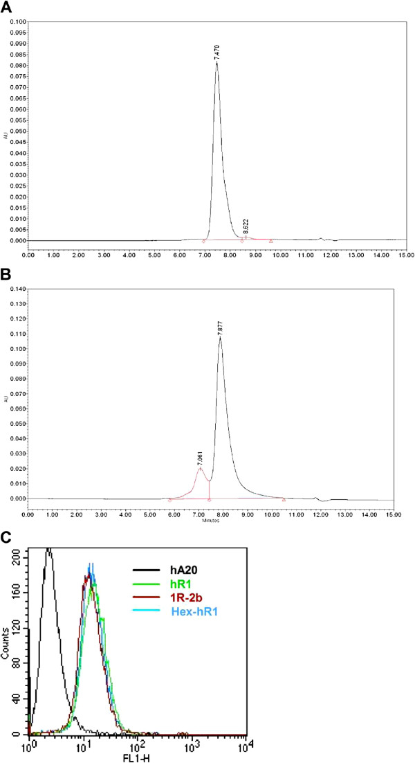
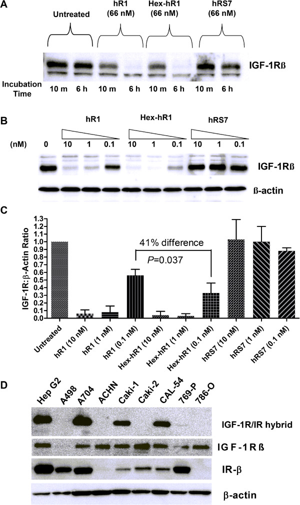
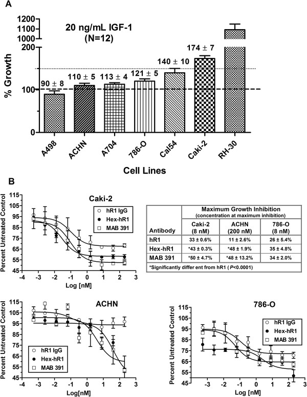
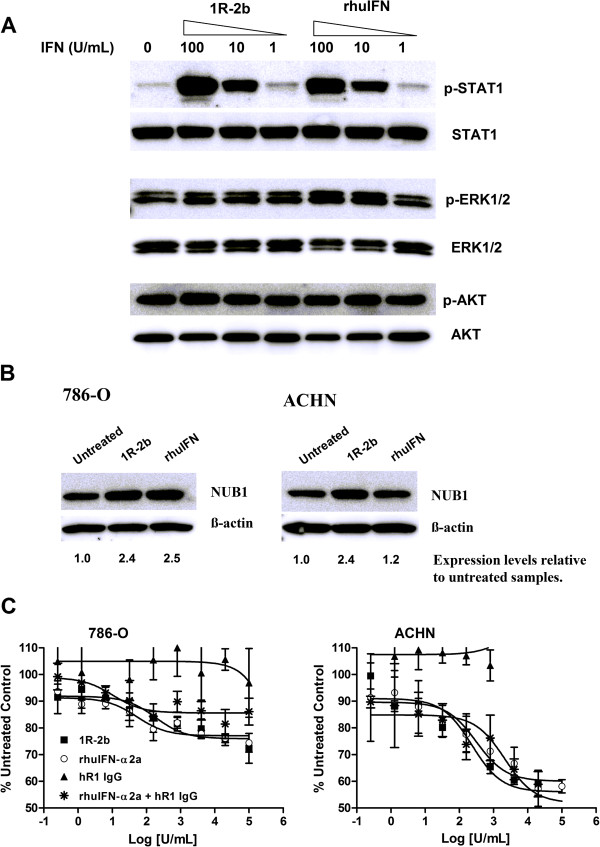
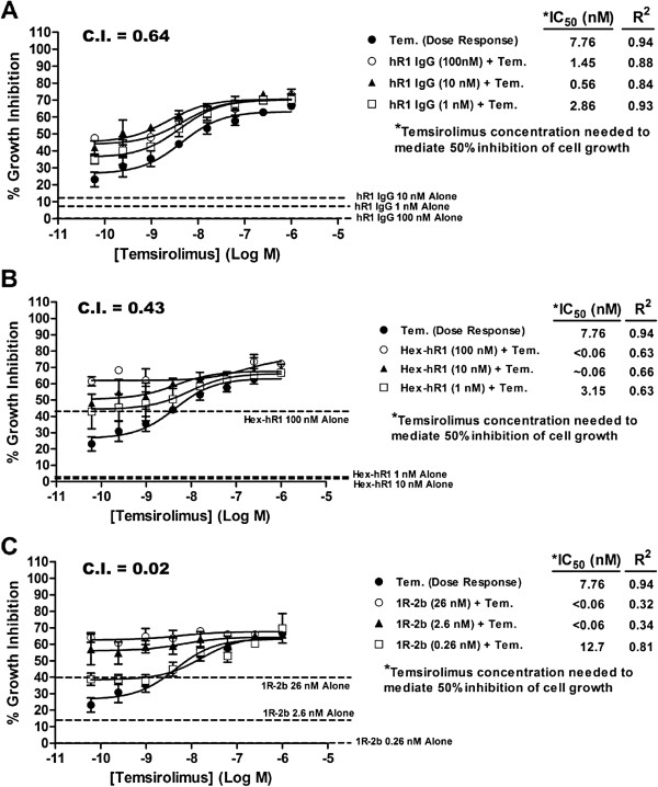
Methods: hR1 was linked by the DOCK-AND-LOCK™ (DNL™) method to four Fabs of hR1, generating Hex-hR1, or to four molecules of interferon-α2b, generating 1R-2b. Eight human RCC cell lines were screened for IGF-1R expression and sensitivity to treatment with hR1 in vitro. Synergy with an mTOR inhibitor, temsirolimus, was tested in a cell line (ACHN) with low sensitivity to hR1.

Results: Hex-hR1 induced the down-regulation of IGF-IR at 10-fold lower concentrations compared to the parental hR1. Sensitivity to growth inhibition mediated by hR1 and Hex-hR1 treatments correlated with IGF-1R expression (higher expression was more sensitive). The potency of 1R-2b to inhibit the in vitro growth of RCC was also demonstrated in two human cell lines, ACHN and 786-O, with EC50-values of 63 and 48 pM, respectively. When combined with temsirolimus, a synergistic growth-inhibition with hR1, Hex-hR1, and 1R-2b was observed in ACHN cells at concentrations as low as 10 nM for hR1, 1 nM for Hex-hR1, and 2.6 nM for 1R-2b.

Conclusions: Both Hex-hR1 and 1R-2b proved to be more potent than parental hR1 in inhibiting growth of RCC in vitro. Synergy was achieved when each of the three hR1-based agents was combined with temsirolimus, suggesting a new approach for treating RCC.


Figures

Figure 1
Figure 1
Characterization of Hex-hR1 and 1R-2b. A 50-μg sample of (A) Hex-hR1 or (B) 1R-2b was run on SE-HPLC as described in Materials and Methods. Histograms show UV absorbance of eluted material versus time. (C) Cell binding of hR1, Hex-hR1, and 1R-2b at equimolar concentrations on ACHN cells by FACS staining. Anti-CD20 antibody, hA20, served as negative control.
Figure 2
Figure 2
IGF-1R expression. (A) ACHN cells were plated overnight in 6-well plates as described in Materials and Methods. Cells were than exposed to hR1, Hex-hR1, or control hRS7 antibodies before being lysed and 20 μg protein from these lysates subjected to SDS-PAGE (4-20%) followed by blotting with an anti-IGF-1Rβ antibody. IGF-1R time-course down-regulation after exposure to constant amount of either hR1 or Hex-hR1. (B) Cells were exposed to indicated amounts of hR1, Hex-hR1, or hRS7 (anti-Trop-2 antibody) for 6 h before being lysed for Western blotting. β-actin served at the loading control. Blot shown is representative of three repeat experiments. (C) Ratio of IGF-1R to β-actin loading control normalized to untreated cells for the various doses of hR1, Hex-hR1 or control hRS7. Data are shown as mean ± standard deviation. Significance set at P < 0.05 for paired t-test of three experiments. (D) Indicated cells were lysed subjected to immunoprecipitation to determine IGF-1R/IR hybrid receptors, as described in Materials and Methods. A 20-μL aliquot from each immounoprecipitation preparation was subjected to gel eletrophoresis and Western blotting. Hep G2 served as the positive control cell line in these experiments [22]. Blot shown is representative of two repeat experiments. To confirm presence of IGF-1R in IP samples, IP blots were probed with anti-IGF-1R antibody. Additionally, cell lysates were subjected to Western analysis and probed with an anti-IRβ antibody to show relative levels of IR in the various cell lines. β-actin served at the loading control.
Figure 3
Figure 3
In vitro sensitivity to anti-IGF-1R treatment. (A) Various RCC cell lines were plated in 96-well plates overnight in SFM media before being stimulate with 20 ng/mL of IGF-1. Cells were incubated a further 96 h before cell viability determined as described in Materials and Methods. Growth relative to cells grown only in SFM are shown in the graph. Large dotted line indicates growth of cells in SFM while smaller dotted line indicates point of 50% increase in growth. RH30 served as a positive control cell line. (B) In vitro cytotoxicity assay was run as described in Materials and Methods and compared hR1, Hex-hR1, and positive control MAB391. After 96-h incubation, growth as a percent of untreated cells is shown in the graphs. The table shows maximum growth-inhibition achieved in the various cell lines with each antibody and the concentration required to achieve that inhibition. Data are shown as mean ± standard deviation.
Figure 4
Figure 4
In vitro potency of 1R-2b. (A) IFN-α mediation of phosphorylation of STAT1, ERK1/2 and AKT in ACHN cells was performed as described in Materials and Methods. Cells were exposed to the indicated amounts of 1R-2b or rhIFN-α2a for 30 min (p-STAT1) or 60 min (p-ERK1/2 and p-AKT). Fold-increase in phosphorylation was calculated relative to total protein loading controls and normalized to untreated cells. (B) NUB1 expression was determined as described in Materials and Methods. ACHN or 786-O cells were exposed to 3000 U/mL of 1R-2b or rhIFN-α2a for 24 h. Up-regulation was determined relative to β-actin loading control and normalized against untreated cells. (C) Growth inhibition was performed as described in Materials and Methods in complete media containing 10% FBS. A dose/response curve was generated with 1R-2b or rhIFN-α2a ranging from 1×10⁵ to 0.26 U/mL. Graphs show growth relative to untreated control and represent the mean ± standard deviation.
Figure 5
Figure 5
Synergistic interaction of temsirolimus with various anti-IGF-1R molecules. Cytotoxicity assays were performed as described in Materials and Methods. A dose/response curve with temsirolimus was made from 1x10⁻⁶ to 6.1x10⁻¹¹ M. To one set of temsirolimus wells was added a constant amount of (A) hR1 or (B) Hex-hR1 at 100 nM, 10 nM, or 1 nM. (C) For 1R-2b, a constant amount of 26, 2.6, or 0.26 nM was added to the wells. After 96 h incubation, growth relative to untreated cells was determined and the amount of temsirolimus required to inhibit cell growth by 50% was calculated for temsirolimus alone or when used in combination with hR1, Hex-hR1, or 1R-2b. C.I. values were determined as described, with a value less than 1 indicative of synergy. Dotted lines indicate growth-inhibition of each anti-IGF-1R agent alone. Data are shown as mean ± standard deviation.

References

    1. American Cancer Society. Cancer Facts and Figures 2012. Atlanta: American Cancer Society; 2012.
    1. Fisher R, Gore M, Larkin J. Current and future systemic treatments for renal cell carcinoma. Semin Cancer Biol. 2013;23:38–45. doi: 10.1016/j.semcancer.2012.06.004. - DOI - PubMed
    1. Aschenbrenner DS. New drug approved for advanced renal cell carcinoma. Am J Nurs. 2012;112:22–23. - PubMed
    1. Gollob JA, Rathmell WK, Richmond TM, Marino CB, Miller EK, Grigson G, Watkins C, Gu L, Peterson BL, Wright JJ. Phase II trial of sorafenib plus interferon alfa-2b as first- or second-line therapy in patients with metastatic renal cell cancer. J Clin Oncol. 2007;25:3288–3295. doi: 10.1200/JCO.2007.10.8613. - DOI - PubMed
    1. Kapoor A, Figlin RA. Targeted inhibition of mammalian target of rapamycin for the treatment of advanced renal cell carcinoma. Cancer. 2009;115:3618–3630. doi: 10.1002/cncr.24409. - DOI - PubMed

Publication types

MeSH terms

Substances