

Localized bi-nasal macular edema in optic chiasmal syndrome
- PMID: 23548317
- PMCID: PMC3759107
- DOI: 10.4103/0301-4738.97079
Localized bi-nasal macular edema in optic chiasmal syndrome
Abstract
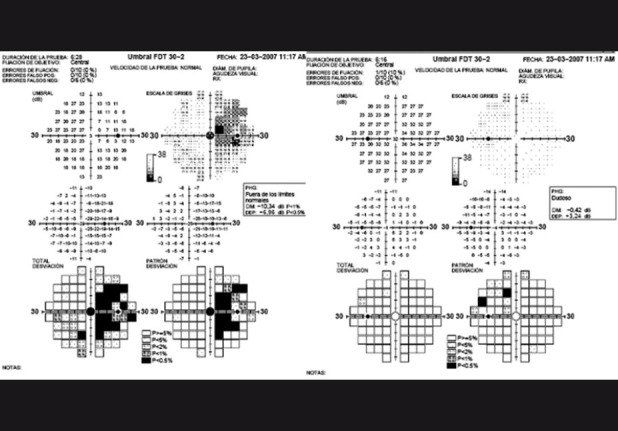
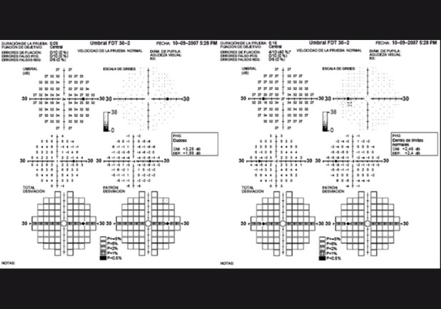
A 28-year-old healthy male complaining of vision loss in his right eye was discovered to have localized bi-nasal macular edema in the presence of a pituitary adenoma. The presence of a junctional scotoma composed by a central scotoma in the right eye associated with superior temporal quadrantanopia in the fellow eye was seen. The pattern detected in the visual field suggested the presence of an expansive mass at the level of the optic chiasm. Optical coherence tomography findings also revealed subtle macular thickness beyond normal in the superior and nasal quadrants of both maculae. This report illustrates the importance of suspecting a pituitary adenoma in the light of uncharacteristic retinal alterations.
Conflict of interest statement
Figures

References
-
- Foroozan R. Chiasmal syndromes. Curr Opin Ophthalmol. 2003;14:325–31. - PubMed
-
- Lederer DE, Schuman JS, Hertzmark E, Heltzer J, Velazques LJ, Fujimoto JG, et al. Analysis of macular volume in normal and glaucomatous eyes using optical coherence tomography. Am J Ophthalmol. 2003;135:838–43. - PubMed
-
- Medeiros FA, Zangwill LM, Bowd C, Vessani RM, Susanna R, Jr, Weinreb RN. Evaluation of retinal nerve fiber layer, optic nerve head, and macular thickness measurements for glaucoma detection using optical coherence tomography. Am J Ophthalmol. 2005;139:44–55. - PubMed
-
- Knox DL, Eagle RC, Jr, Green WR. Optic nerve hydropic axonal degeneration and blocked retrograde axoplasmic transport: Histopathologic features in human high-pressure secondary glaucoma. Arch Ophthalmol. 2007;125:347–53. - PubMed
Publication types
MeSH terms
LinkOut - more resources
Full Text Sources
Other Literature Sources
