. 2013 May 24;288(21):15380-9.
doi: 10.1074/jbc.M112.427765. Epub 2013 Apr 2.

Endomembrane H-Ras controls vascular endothelial growth factor-induced nitric-oxide synthase-mediated endothelial cell migration

Affiliations

Endomembrane H-Ras controls vascular endothelial growth factor-induced nitric-oxide synthase-mediated endothelial cell migration

Dagmar J Haeussler et al. J Biol Chem. .

Abstract

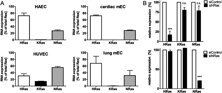
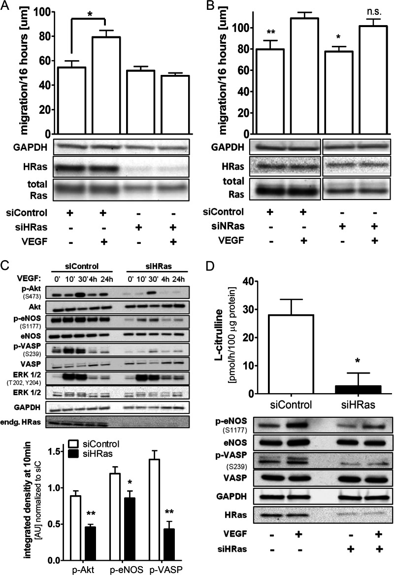
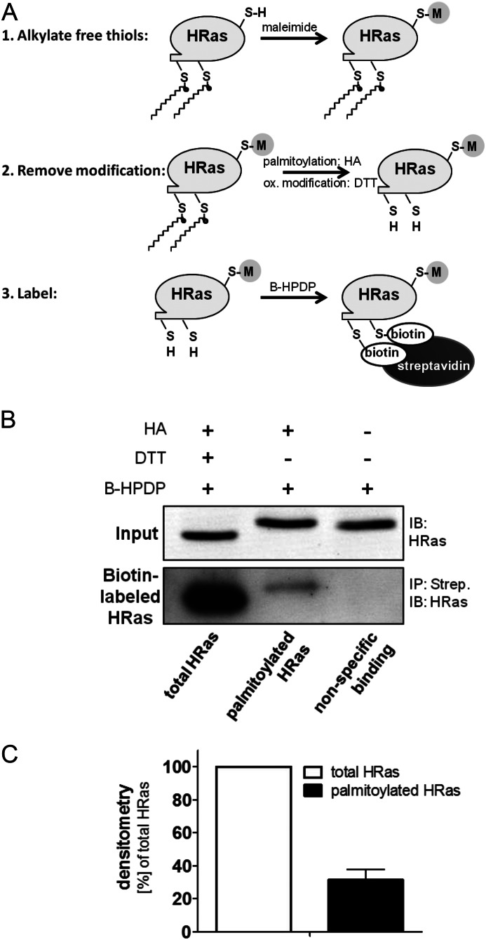
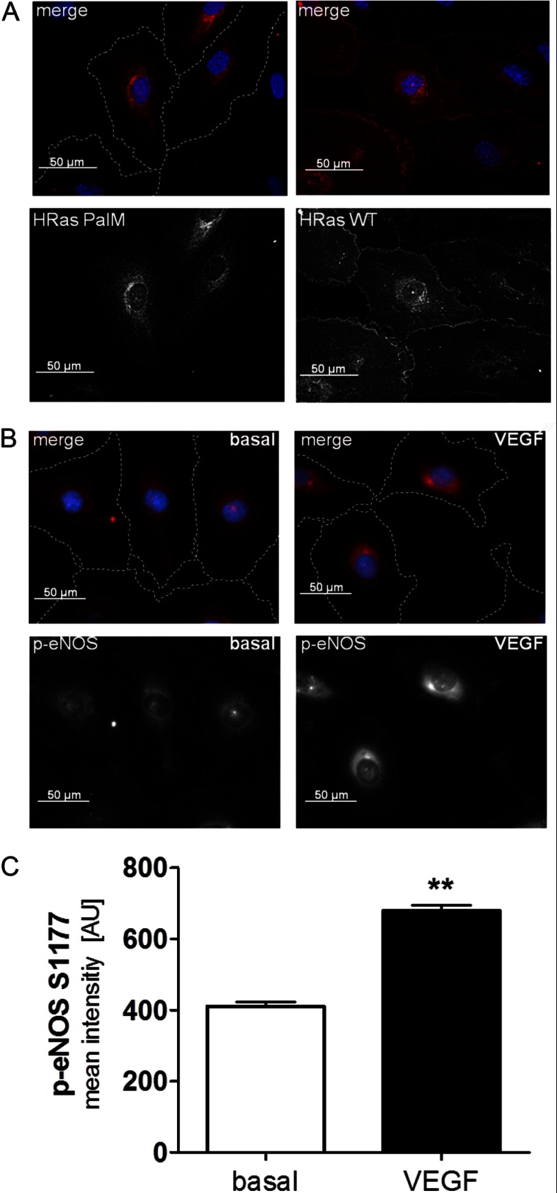
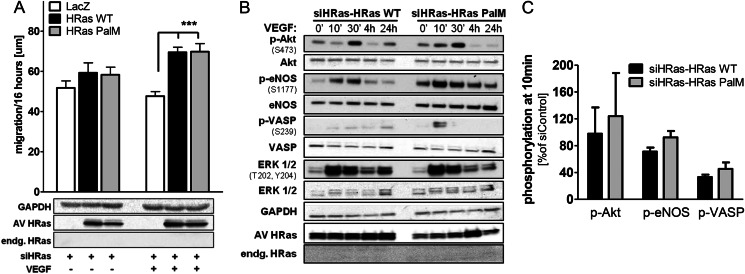
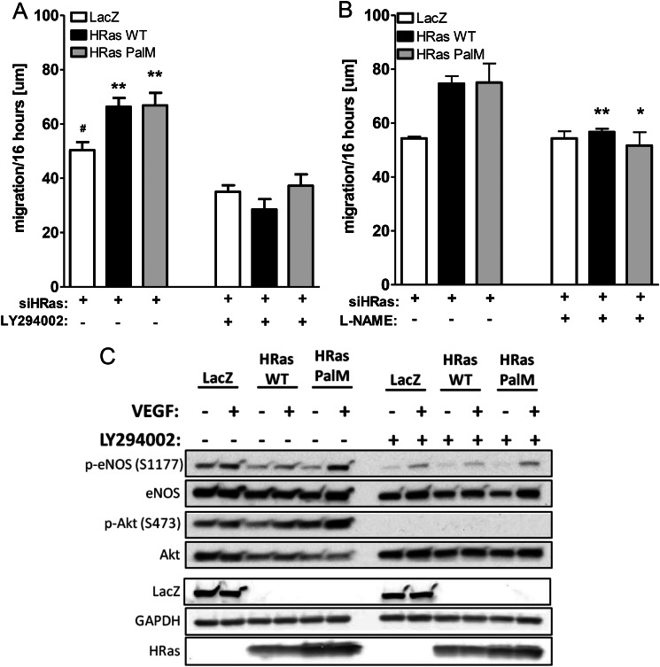
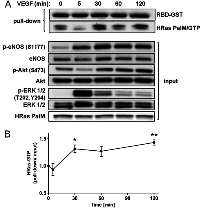
We demonstrate for the first time that endomembrane-delimited H-Ras mediates VEGF-induced activation of endothelial nitric-oxide synthase (eNOS) and migratory response of human endothelial cells. Using thiol labeling strategies and immunofluorescent cell staining, we found that only 31% of total H-Ras is S-palmitoylated, tethering the small GTPase to the plasma membrane but leaving the function of the large majority of endomembrane-localized H-Ras unexplained. Knockdown of H-Ras blocked VEGF-induced PI3K-dependent Akt (Ser-473) and eNOS (Ser-1177) phosphorylation and nitric oxide-dependent cell migration, demonstrating the essential role of H-Ras. Activation of endogenous H-Ras led to recruitment and phosphorylation of eNOS at endomembranes. The loss of migratory response in cells lacking endogenous H-Ras was fully restored by modest overexpression of an endomembrane-delimited H-Ras palmitoylation mutant. These studies define a newly recognized role for endomembrane-localized H-Ras in mediating nitric oxide-dependent proangiogenic signaling.

Keywords: Angiogenesis; Cell Migration; Endomembrane; H-Ras; Nitric Oxide; Nitric-oxide Synthase; Protein Palmitoylation; Vascular Endothelial Growth Factor (VEGF).


Figures

FIGURE 1.
FIGURE 1.
Relative expression level of the three major Ras isoforms in HAECs and other types of ECs. A, relative expression levels of Ras isoforms in untreated HAECs, HUVECs, cardiac mouse ECs, and lung mouse ECs normalized to β-actin were measured with TaqMan quantitative PCR of mRNA expression levels. n ≥ 3. Error bars, S.E. B, efficacy of H-Ras and N-Ras knockdown. Endogenous H-Ras mRNA was knocked down by targeting the 3′-untranslated region. Relative expression levels of HAECs treated with siControl or siH-Ras are shown. Two-way ANOVA and Bonferroni's post-test were used (n ≥ 4). Error bars, S.E. ***, p < 0.001, siControl versus siH-Ras; n.s., not significant.
FIGURE 2.
FIGURE 2.
VEGF-induced EC migration is H-Ras-dependent. A, knockdown of endogenous H-Ras blocks EC migratory response to VEGF. After overnight starvation, a wound healing assay was performed with HAECs using VEGF (50 ng/ml) as a stimulus. One-way ANOVA and Dunnett's post-test were used (n ≥ 4). Error bars, S.E. *, p < 0.05. B, knockdown of endogenous N-Ras has no significant effect on EC migration in response to VEGF. A wound healing assay was performed as described in A. All depicted bands were on the same membrane and only cut for visual clarity. One-way ANOVA and Dunnett's post-test were used (n ≥ 3). Error bars, S.E. *, p < 0.05; **, p < 0.01. VEGF stimulation versus no stimulation; n.s., not significant, VEGF-stimulated siNRas versus VEGF-stimulated siControl. C, knockdown of endogenous H-Ras in HAECs resulted in loss of the key proangiogenic signaling cascade Akt/eNOS, including downstream NO/PKG effector VASP in response to VEGF. HAECs treated with either siControl or siH-Ras were stimulated after overnight starvation for 0 min, 10 min, 30 min, 4 h, and 24 h with VEGF (50 ng/ml). Densitometric quantification of HAECs treated with either siControl or siH-Ras at 10 min of VEGF stimulation. All values were standardized to GAPDH as a loading control and normalized to siControl at 0 min. Unpaired Student's t test was used (n ≥ 3). Error bars, S.E. **, p < 0.01; *, p < 0.05. D, VEGF-stimulated eNOS activity (Ser-1177 phosphorylation) was reduced by knockdown of endogenous H-Ras. H-Ras-depleted HAECs were stimulated for 30 min with VEGF (50 ng/ml) and lysed. Part of the lysate was processed for Western blotting (bottom), and eNOS activity (top) was measured in vitro by quantifying 14C-labeled l-arginine to 14C-labeled l-citrulline conversion using a phosphor imager system. Band intensity for basal level was subtracted from VEGF-stimulated eNOS activity. Unpaired Student's t test was used (n = 4). Error bars, S.E. *, p < 0.05.
FIGURE 3.
FIGURE 3.
The majority of H-Ras is not palmitoylated. A, hydroxylamine (HA)-dependent biotin switch is a reversed labeling technique for palmitoylated cysteines. First, free thiols (RSH) are alkylated by maleimide. Then thioester bonds, including palmitoylation, are hydrolyzed by HA, whereas reversible oxidative (ox) modifications are removed by DTT. The freed cysteines are subsequently labeled by thiol-reactive HPDP-Biotin (B-HPDP), and biotinylated protein is enriched using streptavidin pull-down. B, palmitoylation of endogenous H-Ras was measured using the HA biotin switch assay. Lysates were treated either with DTT and HA to label total H-Ras or with HA only to specifically remove S-palmitoylation and label freed cysteines with thiol-reactive HPDP-Biotin. The pull-down on streptavidin-agarose (Strep) was separated by SDS-PAGE and immunoblotted for H-Ras with an isoform-specific antibody. IB, immunoblot; IP, immunoprecipitation. C, densitometry was performed for streptavidin pull-down (n = 3). Error bars, S.E.
FIGURE 4.
FIGURE 4.
Subcellular distribution of H-Ras and phospho-eNOS after VEGF stimulation. A, left, the palmitoylation-deficient H-Ras PalM (C181S/C184S) localizes predominantly to endomembranes. Right, adenoviral H-Ras WT localizes to the plasma membrane and the perinuclear region in EC. For detection of the overexpressed H-Ras WT and PalM, an antibody against the XPRESS affinity tag was used. Pictures were taken by confocal microscopy. Cell borders are outlined with a white dashed line for H-Ras PalM. B, eNOS phosphorylation at serine Ser-1177 is increased in the perinuclear region after 30-min VEGF stimulation. HAECs were starved overnight, treated with VEGF (30 min, 50 ng/ml), and subsequently fixed and stained for eNOS (phospho S1177, red) and cell nuclei (DAPI, blue). Staining was performed as described under “Materials and Methods.” Pictures were taken at ×400, using epifluorescence. Cell borders are outlined with a white dashed line in the merged pictures. C, mean fluorescent intensity of staining, expressed in arbitrary units (AU), for eNOS phosphorylation at serine 1177 was measured in the perinuclear region in 35–50 cells/experiment under the same conditions using NIS-Elements software. Unpaired Student's t test was used (n = 2). Error bars, S.E. **, p < 0.01.
FIGURE 5.
FIGURE 5.
Endomembrane H-Ras is mediating VEGF-induced migration by activating the PI3K/Akt/eNOS signaling cascade. A, in H-Ras-depleted ECs, reconstitution of endogenous H-Ras with either adenoviral H-Ras WT or endomembrane H-Ras PalM resulted in recovery of migration in response to VEGF. After overnight starvation, a wound healing assay was performed with H-Ras-depleted HAECs using VEGF (50 ng/ml) as a stimulus. Two-way ANOVA and Bonferroni's post-test were used (n ≥ 5). Error bars, S.E. ***, p < 0.001. B, reconstitution of endogenous H-Ras with either adenoviral H-Ras WT or H-Ras PalM resulted in partial recovery of the key proangiogenic signaling cascade Akt/eNOS in response to VEGF. HAECs treated with siH-Ras and reconstituted with H-Ras WT or PalM were stimulated after overnight starvation for 0 min, 10 min, 30 min, 4 h, and 24 h with VEGF (50 ng/ml). C, densitometric quantification of HAECs treated with siH-Ras and reconstituted with H-Ras WT or H-Ras PalM at 10-min VEGF stimulation. All values were standardized to GAPDH as a loading control and normalized to siC-LacZ at 0 min. n ≥ 3. Error bars, S.E.
FIGURE 6.
FIGURE 6.
Endomembrane H-Ras activates NO production by activating the PI3K/Akt/eNOS signaling cascade. A, H-Ras-mediated EC migration was inhibited by inhibition of PI3K. H-Ras depleted and reconstituted with either LacZ, H-Ras WT, or H-Ras PalM HAECs were stimulated with VEGF (50 ng/ml), and an EC wound healing assay was performed. ECs were treated with either DMSO or the PI3K inhibitor LY294002 (5 μm) during migration. One-way ANOVA and Dunnett's post-test were used (n ≥ 4). Error bars, S.E. **, p < 0.01, treatment versus no treatment; *, p < 0.05, reconstitution with LacZ versus H-Ras WT or PalM. B, H-Ras-mediated EC migration was reduced by inhibiting eNOS with l-NAME. The wound healing assay was performed as described above, and cells were treated with l-NAME (1 mm) for the duration of the assay. Unpaired Student's t test was used (n ≥ 3). Error bars, S.E. **, p < 0.01; *, p < 0.05, treatment versus no treatment. C, H-Ras-mediated proangiogenic signaling was reduced by inhibition of PI3K. H-Ras-depleted and -reconstituted HAECs were preincubated with LY294002 (10 μm) and then stimulated with VEGF (50 ng/ml, 30 min). A representative Western blot is shown.
FIGURE 7.
FIGURE 7.
Endomembrane H-Ras is activated by VEGF stimulation. A, HAECs depleted of endogenous H-Ras and reconstituted with H-Ras PalM were starved overnight and treated for increasing periods of time with VEGF (50 ng/ml). Cells were harvested in RBD-GST-containing lysis buffer, and active H-Ras (H-Ras/GTP) was pulled down using glutathione-agarose beads. Coomassie-stained RBD-GST was shown to demonstrate equal loading of RBD-GST. Representative blots of pull-down and input were shown. B, pulled down H-Ras PalM quantified using ImageJ and normalized to H-Ras input. One-way ANOVA and Dunnett's post-test were used (n ≥ 4). Error bars, S.E. *, p < 0.05; **, p < 0.01.

Similar articles

Cited by

References

    1. Castellano E., Downward J. (2011) RAS interaction with PI3K. More than just another effector pathway. Genes Cancer 2, 261–274 - PMC - PubMed
    1. Hancock J. F. (2003) Ras proteins. Different signals from different locations. Nat. Rev. Mol. Cell Biol. 4, 373–384 - PubMed
    1. Ahearn I. M., Haigis K., Bar-Sagi D., Philips M. R. (2012) Regulating the regulator. Post-translational modification of RAS. Nat. Rev. Mol. Cell Biol. 13, 39–51 - PMC - PubMed
    1. Swarthout J. T., Lobo S., Farh L., Croke M. R., Greentree W. K., Deschenes R. J., Linder M. E. (2005) DHHC9 and GCP16 constitute a human protein fatty acyltransferase with specificity for H- and N-Ras. J. Biol. Chem. 280, 31141–31148 - PubMed
    1. Henis Y. I., Hancock J. F., Prior I. A. (2009) Ras acylation, compartmentalization, and signaling nanoclusters (Review). Mol. Membr. Biol. 26, 80–92 - PMC - PubMed

Publication types

MeSH terms

Substances