. 2013 May 17;288(20):14647-14656.
doi: 10.1074/jbc.M112.439893. Epub 2013 Apr 2.

Matrix metalloproteinase Mmp-1a is dispensable for normal growth and fertility in mice and promotes lung cancer progression by modulating inflammatory responses

Affiliations

Matrix metalloproteinase Mmp-1a is dispensable for normal growth and fertility in mice and promotes lung cancer progression by modulating inflammatory responses

Miriam Fanjul-Fernández et al. J Biol Chem. .

Erratum in

Abstract

Human MMP-1 is a matrix metalloproteinase repeatedly associated with many pathological conditions, including cancer. Thus, MMP1 overexpression is a poor prognosis marker in a variety of advanced cancers, including colorectal, breast, and lung carcinomas. Moreover, MMP-1 plays a key role in the metastatic behavior of melanoma, breast, and prostate cancer cells. However, functional and mechanistic studies on the relevance of MMP-1 in cancer have been hampered by the absence of an in vivo model. In this work, we have generated mice deficient in Mmp1a, the murine ortholog of human MMP1. Mmp1a(-/-) mice are viable and fertile and do not exhibit obvious abnormalities, which has facilitated studies of cancer susceptibility. These studies have shown a decreased susceptibility to develop lung carcinomas induced by chemical carcinogens in Mmp1a(-/-) mice. Histopathological analysis indicated that tumors generated in Mmp1a(-/-) mice are smaller than those of wild-type mice, consistently with the idea that the absence of Mmp-1a hampers tumor progression. Proteomic analysis revealed decreased levels of chitinase-3-like 3 and accumulation of the receptor for advanced glycation end-products and its ligand S100A8 in lung samples from Mmp1a(-/-) mice compared with those from wild-type. These findings suggest that Mmp-1a could play a role in tumor progression by modulating the polarization of a Th1/Th2 inflammatory response to chemical carcinogens. On the basis of these results, we propose that Mmp1a knock-out mice provide an excellent in vivo model for the functional analysis of human MMP-1 in both physiological and pathological conditions.

Keywords: Carcinogenesis; Degradome; Inflammation; Invasion; Metastasis; Protease.


Figures

FIGURE 1.
FIGURE 1.
Targeted disruption of mouse Mmp1a gene. A, restriction maps of the Mmp1a gene region of interest (top), the targeting construct (center), and the mutant locus after homologous recombination (bottom). B, EcoRI Southern blot analysis of Mmp1a+/+, Mmp1a+/−, and Mmp1a−/− mice. C, detection of Mmp1a mRNA in placenta by Northern blot analysis.
FIGURE 2.
FIGURE 2.
Mmp1a relevance on cancer susceptibility in mice. A, mRNA expression levels in tissues from wild-type mice subjected to different experimental protocols. MMTV-PyMT, mouse mammary tumor virus-polyoma middle T antigen; K14-HPV16, keratin 14-human papillomavirus type 16; MCA, 3-methyl-cholanthrene; DMBA, 9,10-dimethyl-1,2-benzanthracene. DEN, N,N-diethylnitrosamine; 4-NQO, 4-nitroquinoline 1-oxide; CCl4, carbon tetrachloride. B, number of total lung tumors per mouse after 8 months of urethane intraperitoneal treatment. C, representative images of lungs from mutant and control mice (n = 15 per group; ***, p < 0.001 by Student's t test.
FIGURE 3.
FIGURE 3.
Phenotypic features of urethane-induced tumors in Mmp1a wild-type and knock-out mice. A, graphic representation of size tumor incidence in males. Knock-out mice show a lower incidence of large tumors (>103 cells). B, inflammatory infiltrate incidence in both sexes. We represent the number of mice which present inflammatory infiltrate foci in their lungs, showing an increased presence of inflammatory foci in lungs from knock-out mice. Inflammatory infiltrates are indicated with arrows, and tumors are shown with arrowheads.
FIGURE 4.
FIGURE 4.
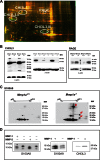
A, difference gel electrophoresis analysis of lung tissue from urethane-treated mice. Overlaying green and red image highlights differences between wild-type and Mmp1a−/− mice. Yellow indicates no change, red spots indicate more abundance in knock-out mice, and green spots more abundance in wild-type mice. Selected proteins are labeled with white circles, and bidimensional validation analysis is shown in supplemental Fig. 1. All differential analyzed spots are listed in supplemental Table 1. B, Western blot analysis extended to other urethane-treated littermates showing the increased CHI3L3 and RAGE levels in wild-type and knock-out mice, respectively, as well as the differential processing of CHI3L3 in wild-type lungs no present in the mutant lungs. Load control is shown at the bottom of each panel. C, Western blot analysis of the RAGE ligand S100A8 showing accumulation of different isoforms of this chemokine in lung from knock-out mice. D, in vitro cleavage assays with human MMP-1. Purified S100A8, S100A9, and CHI3L3 (1 μg) were incubated with 100 ng of activated MMP-1, which resulted in specific cleavage of S100A8 (*), but not S100A9 and CHI3L3. Recombinant MMP-2 was used as a positive control of S100A8 cleavage.
FIGURE 5.
FIGURE 5.
Analysis of cytokine levels in lung from Mmp1a wild-type and knock-out mice treated with urethane. A, cytokine levels measured in lung mice after 8 months of urethane intraperitoneal treatment. B, measurement of IFN-γ and IL-17 in the lungs of mutant and control mice at several time points along the urethane carcinogenesis model. C, levels of Th1 cytokines in lungs from two different groups of Mmp1a-deficient mice separated on the basis of their inflammatory phenotype. Mmp1a−/− group A (Mmp1a −/− A) represents those mutant mice showing a notably high number of inflammatory infiltrate foci in the lungs, whereas Mmp1a−/− group B (Mmp1a−/− B) represents those mutant mice with no overt infiltrate foci in their lungs. *, p < 0.01; **, p < 0.001 by Student's t test.

References

    1. Fanjul-Fernández M., Folgueras A. R., Cabrera S., López-Otín C. (2010) Matrix metalloproteinases: evolution, gene regulation and functional analysis in mouse models. Biochim. Biophys. Acta 1803, 3–19 - PubMed
    1. Kessenbrock K., Plaks V., Werb Z. (2010) Matrix metalloproteinases: regulators of the tumor microenvironment. Cell 141, 52–67 - PMC - PubMed
    1. Coussens L. M., Fingleton B., Matrisian L. M. (2002) Matrix metalloproteinase inhibitors and cancer: trials and tribulations. Science 295, 2387–2392 - PubMed
    1. Overall C. M., López-Otín C. (2002) Strategies for MMP inhibition in cancer: innovations for the post-trial era. Nat. Rev. Cancer 2, 657–672 - PubMed
    1. Egeblad M., Werb Z. (2002) New functions for the matrix metalloproteinases in cancer progression. Nat. Rev. Cancer 2, 161–174 - PubMed

Publication types

LinkOut - more resources