. 2013 Apr;123(4):1501-12.
doi: 10.1172/JCI66576. Epub 2013 Mar 15.

Mycolactone activation of Wiskott-Aldrich syndrome proteins underpins Buruli ulcer formation

Affiliations

Mycolactone activation of Wiskott-Aldrich syndrome proteins underpins Buruli ulcer formation

Laure Guenin-Macé et al. J Clin Invest. 2013 Apr.

Abstract

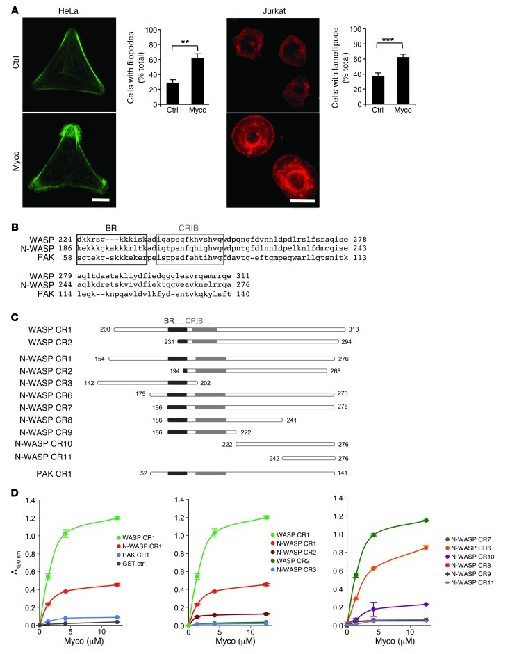
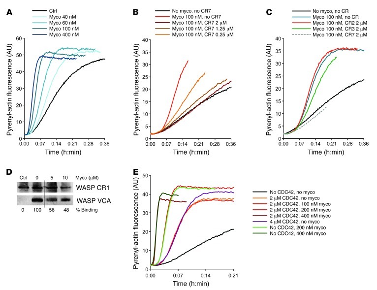
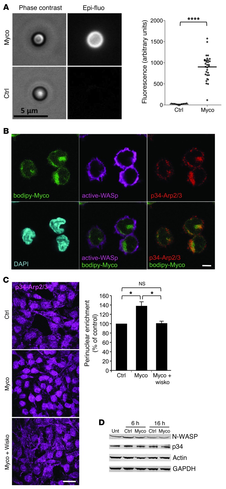
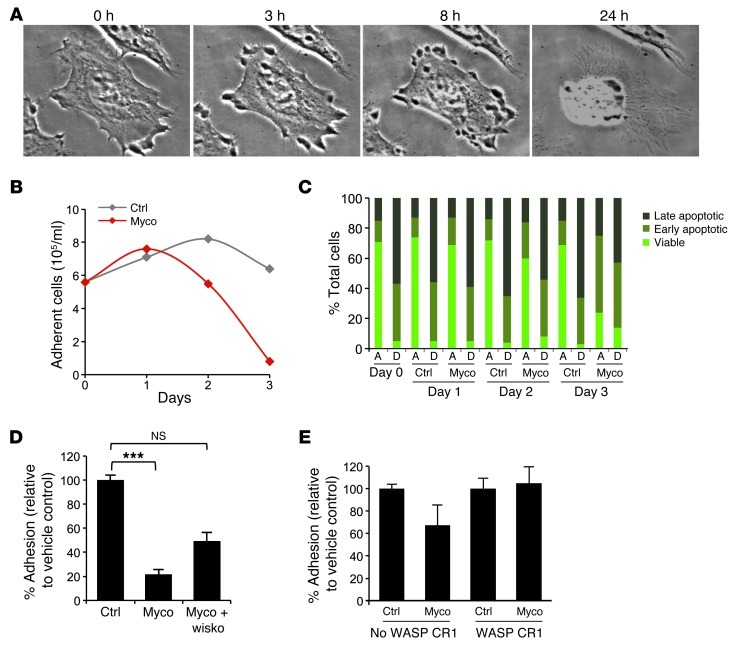
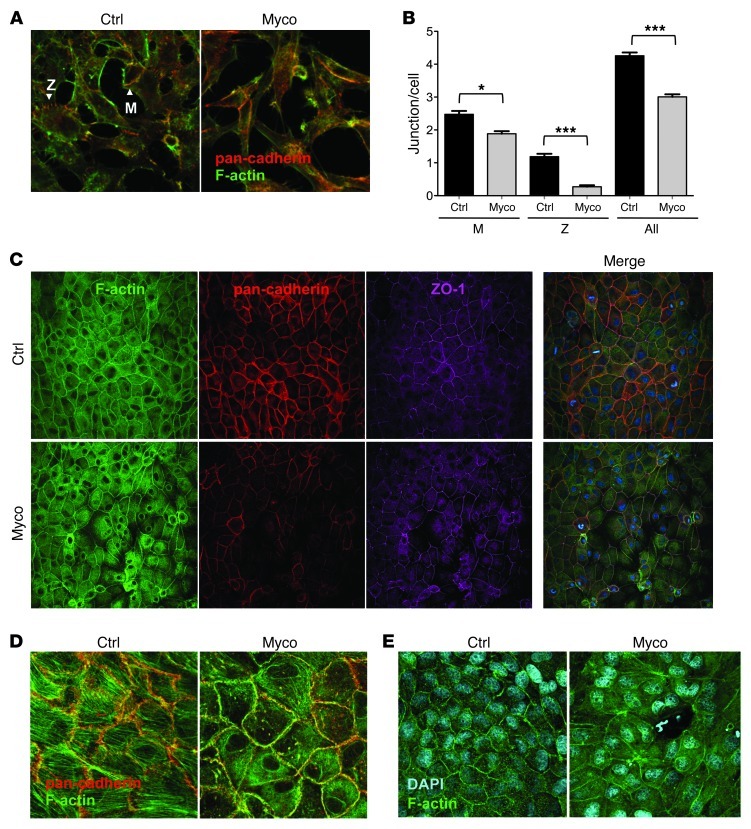
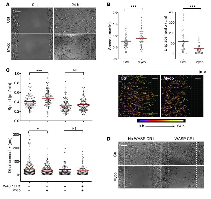
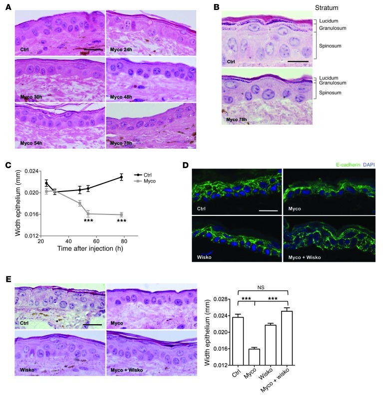
Mycolactone is a diffusible lipid secreted by the human pathogen Mycobacterium ulcerans, which induces the formation of open skin lesions referred to as Buruli ulcers. Here, we show that mycolactone operates by hijacking the Wiskott-Aldrich syndrome protein (WASP) family of actin-nucleating factors. By disrupting WASP autoinhibition, mycolactone leads to uncontrolled activation of ARP2/3-mediated assembly of actin in the cytoplasm. In epithelial cells, mycolactone-induced stimulation of ARP2/3 concentrated in the perinuclear region, resulting in defective cell adhesion and directional migration. In vivo injection of mycolactone into mouse ears consistently altered the junctional organization and stratification of keratinocytes, leading to epidermal thinning, followed by rupture. This degradation process was efficiently suppressed by coadministration of the N-WASP inhibitor wiskostatin. These results elucidate the molecular basis of mycolactone activity and provide a mechanism for Buruli ulcer pathogenesis. Our findings should allow for the rationale design of competitive inhibitors of mycolactone binding to N-WASP, with anti-Buruli ulcer therapeutic potential.


Figures

Figure 1
Figure 1. Mycolactone binds to WASP/N-WASP with high affinity and specificity.
(A) HeLa cells were plated on Y-shaped, fibronectin-coated micropatterns, then exposed to 20 nM mycolactone for 30 minutes. Jurkat T cells were exposed to 20 nM mycolactone for 5 minutes, then spun onto poly-l-lysine–coated glass slides. Actin structures were visualized by phalloidin in cells exposed to mycolactone (Myco) or solvent as control (Ctrl). Scale bars: 10 μm. Graphs show mean cell proportions (± SEM) on more than 50 cells from at least 2 independent experiments. **P < 0.01, ***P < 0.001, unpaired 2-tailed t test. (B) Amino acid sequences of the GBD of WASP, N-WASP, and PAK, with boxes outlining the BR and CRIB motifs. (C) Constructs used in this study. (D) Binding of biotinylated mycolactone to these constructs in ELISA. Data are mean ± SD A490 nm of duplicates.
Figure 2
Figure 2. Mycolactone binding to N-WASP potentiates its actin polymerization activity.
(A) Effect of increasing concentrations of mycolactone on N-WASP–dependent polymerization of pyrenyl actin. Equivalent volumes of solvent were used as control. (B) CR7 dose-dependently inhibited mycolactone-induced N-WASP activation. (C) Effect of CR2, CR3, and CR7 on mycolactone-induced N-WASP activation. (D) Mycolactone dose-dependently displaced CR1-bound VCA. Silver staining of WASP CR1 and VCA, after incubation of GST-fused CR1 (immobilized on glutathione-sepharose beads) with VCA in the presence of increasing amounts of mycolactone, and analysis of bead-bound products by gel electrophoresis. No VCA served as control. Lanes were run on the same gel but were noncontiguous (black line). (E) Differential activity of mycolactone and CDC42-GTP (CDC42) on N-WASP–dependent actin polymerization.
Figure 3
Figure 3. Mycolactone activates cellular WASPs.
(A) Streptavidin-latex beads were coated with biotinylated mycolactone or solvent control, then incubated with N-WASP and placed in Jurkat T cell extracts supplemented with ATP, MgCl2, and Alexa Fluor 488–labeled actin monomers for 2 hours. Representative beads are shown in phase-contrast and fluorescence. Mean fluorescence signal of more than 30 beads from 3 independent experiments are compared. ****P < 0.0001, unpaired 2-tailed t test, Welch corrected. (B) Jurkat cells were treated with a mycolactone fluorescent derivative (bodipy-Myco) for 1 hour, then processed for immunofluorescence with antibody binding the open form of WASP (active-WASP) and antibody recognizing p34-ARP2/3. Each image corresponds to a single confocal plane. 8 of 11 randomly picked cells had a Pearson coefficient greater than 0.5 (calculated on a z stack), indicative of Bodipy-mycolactone and active WASP colocalization. (C) Differential ARP2/3 complex recruitment in HeLa cells treated with vehicle (control), 20 nM mycolactone for 4 hours, or mycolactone plus 1 μM wiskostatin (Wisko). Representative immunofluorescence images and integrated p34-ARP2/3 intensities (n > 50) in the perinuclear region (see Methods) are shown. Perinuclear enrichment was calculated as mean ± SEM intensity and presented relative to control. *P < 0.05, ANOVA with Dunn post-test. (D) Western blot analysis of N-WASP, p34-ARP2/3, and actin expression in HeLa cells exposed to 20 nM mycolactone for 6 or 16 hours, compared with vehicle-treated control cells and untreated cells (Unt). GAPDH served as an internal control. Scale bars: 5 μm (A and B); 25 μm (C).
Figure 4
Figure 4. Effect of mycolactone on epithelial cell adhesion.
(A) Phase-contrast images of a HeLa cell treated with 20 nM mycolactone for 0, 3, 8, and 24 hours. Original magnification, ×63. (B) Mean number of adherent HeLa cells after treatment with 20 nM mycolactone or vehicle control for up to 3 days. (C) Proportion of cells undergoing early and late apoptosis, in the adherent (A) and detached (D) fractions of HeLa cells treated with 20 nM mycolactone or solvent as control. Data are mean percentages on triplicates. (D) Adhesion of HeLa cells, as measured by calcein-AM assay, after 4 hours of treatment with vehicle control, 20 nM mycolactone, or mycolactone in the presence of 1 μM wiskostatin. ***P < 0.001, Kruskal Wallis with Dunn post-test. (E) Adhesion of HeLa cells transfected with an expression vector encoding WASP CR1 or with an empty vector (no WASP CR1) 24 hours prior to treatment with vehicle control or 10 nM mycolactone for 16 hours.
Figure 5
Figure 5. Effect of mycolactone on epithelial cell-cell contacts.
(A) HeLa cell network after 16 hours of treatment with solvent control or 20 nM mycolactone. Characteristic adhesion zipper (Z) and mature (M) junctions are indicated. (B) Effect of mycolactone (20 nM for 16 hours) on the number of intercellular junctions. Data are mean ± SEM junctions per cell measured on more than 200 cells. *P < 0.05, ***P < 0.001, Kruskal Wallis with Dunn post-test. (C) MDCK cell monolayer after 16 hours of treatment with solvent control or 200 nM mycolactone. (D) Confocal image of a basal section. (E) Rupture in the epithelial layer after treatment with 50 nM mycolactone for 16 hours. Original magnification, ×40 (A and E); ×20 (C); ×63 (D).
Figure 6
Figure 6. Mycolactone impairs directed migration of epithelial cells.
(A) Representative image of the wound area after 24 hours of cell migration in the presence of 25 nM mycolactone or methanol (control). Dashed line denotes wound edges at 0 hours. (B) Mean ± SEM cell speed and x displacement (n > 170), and time-dependent representation of cell trajectories. ***P < 0.001, Mann-Whitney test. (C) Mean ± SEM speed and x displacement of more than 200 HeLa cells transfected with an expression vector encoding WASP CR1 24 hours before assessment of cell migration in the presence or absence of 25 nM mycolactone. Controls are cells transfected with empty vector and treated with vehicle. *P < 0.05, ***P < 0.001, Kruskal Wallis with Dunn post-test. (D) Representative images of the wound area after 24 hours of cell migration. HeLa cells were transfected with an expression vector encoding WASP CR1, or an empty vector (no WASP CR1) 24 hours prior to wound formation and cell migration in the presence of 25 nM mycolactone or vehicle control. Dashed lines denote wound edges at 0 hours. Wound-healing assays were repeated twice with similar results. Scale bars: 100 μm (A and D); 50 μm (B).
Figure 7
Figure 7. Remodeling of the skin epidermis after mycolactone administration.
(A) H&E-stained sections of ear skin 24, 30, 48, 54, and 78 hours after intradermal injection of vehicle control or 7 nmol (5 μg) mycolactone. (B) Higher-magnification images of control and 78-hour mycolactone sections, showing thinning of all epidermal layers and loss of stratum granulosum. (C) Evolution of epidermal width at the site of injection of mycolactone or vehicle control. Data are mean ± SEM from 10 measurements per mouse in 3 mice. ***P < 0.001, Kruskal Wallis with Dunn post-test. (D and E) E-cadherin staining (D) and H&E staining and mean width (E) of the ear epidermis 78 hours after intradermal injection of 7 nmol mycolactone, 14 nmol wiskostatin, or both compared with vehicle control injection. Shown are representative images from 3 mice per group. Data are mean ± SEM from 10 measurements per mouse in 3 mice. ***P < 0.001, Kruskal Wallis with Dunn post-test. Scale bars: 20 μm (A, D, and E); 10 μm (B).

References

    1. Demangel C, Stinear TP, Cole ST. Buruli ulcer: reductive evolution enhances pathogenicity of Mycobacterium ulcerans. Nat Rev Microbiol. 2009;7(1):50–60. doi: 10.1038/nrmicro2077. - DOI - PubMed
    1. Merritt RW, et al. Ecology and transmission of Buruli ulcer disease: a systematic review. PLoS Negl Trop Dis. 2010;4(12):e911. doi: 10.1371/journal.pntd.0000911. - DOI - PMC - PubMed
    1. Guarner J, et al. Histopathologic features of Mycobacterium ulcerans infection. Emerg Infect Dis. 2003;9(6):651–656. doi: 10.3201/eid0906.020485. - DOI - PMC - PubMed
    1. George KM, et al. Mycolactone: a polyketide toxin from Mycobacterium ulcerans required for virulence. Science. 1999;283(5403):854–857. doi: 10.1126/science.283.5403.854. - DOI - PubMed
    1. Hong H, Demangel C, Pidot SJ, Leadlay PF, Stinear T. Mycolactones: immunosuppressive and cytotoxic polyketides produced by aquatic mycobacteria. Nat Prod Rep. 2008;25(3):447–454. doi: 10.1039/b803101k. - DOI - PMC - PubMed

Publication types

MeSH terms