. 2013 Apr;123(4):1784-97.
doi: 10.1172/JCI60415. Epub 2013 Mar 15.

Myeloid cell-specific serine palmitoyltransferase subunit 2 haploinsufficiency reduces murine atherosclerosis

Affiliations

Myeloid cell-specific serine palmitoyltransferase subunit 2 haploinsufficiency reduces murine atherosclerosis

Mahua Chakraborty et al. J Clin Invest. 2013 Apr.

Erratum in

  • J Clin Invest. 2013 May;123(5):2332

Abstract

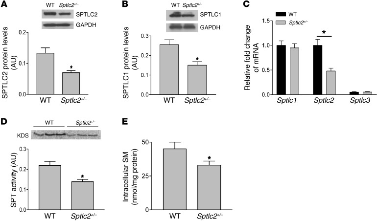
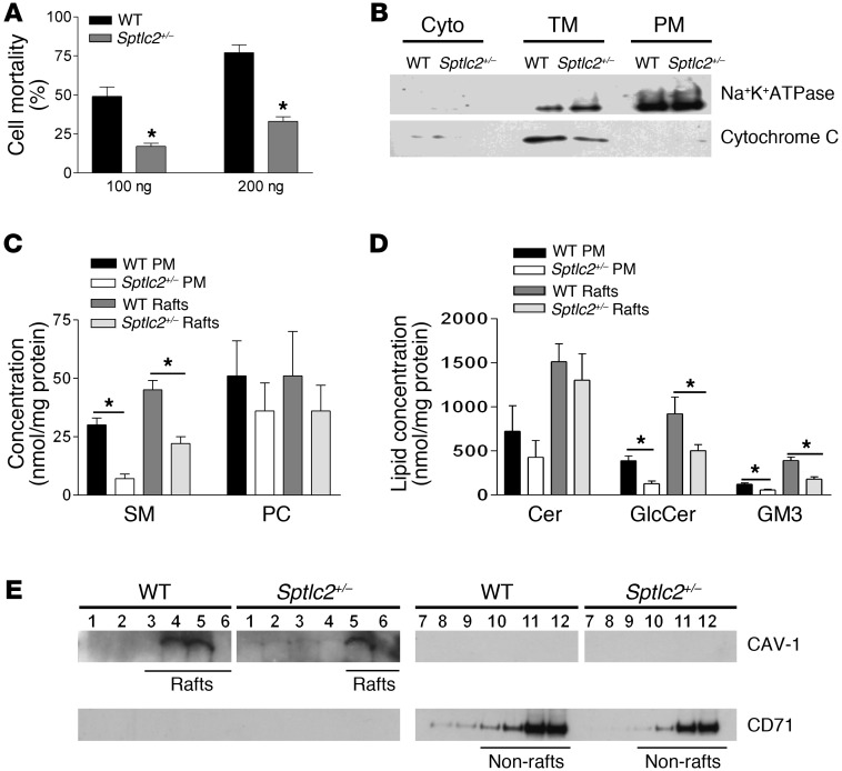
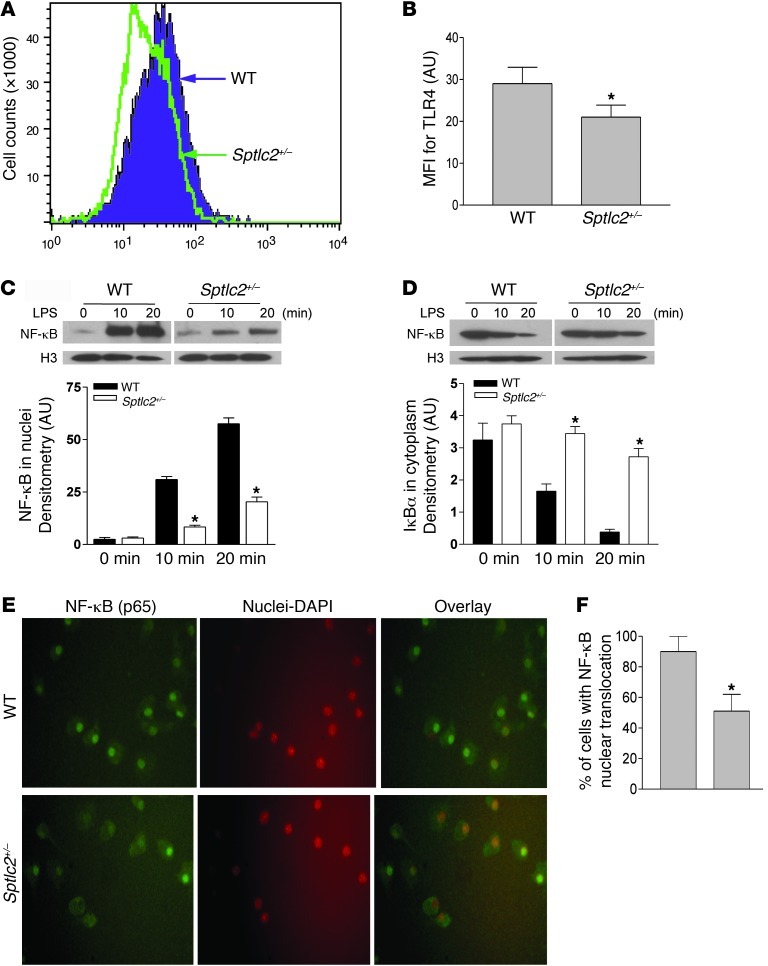
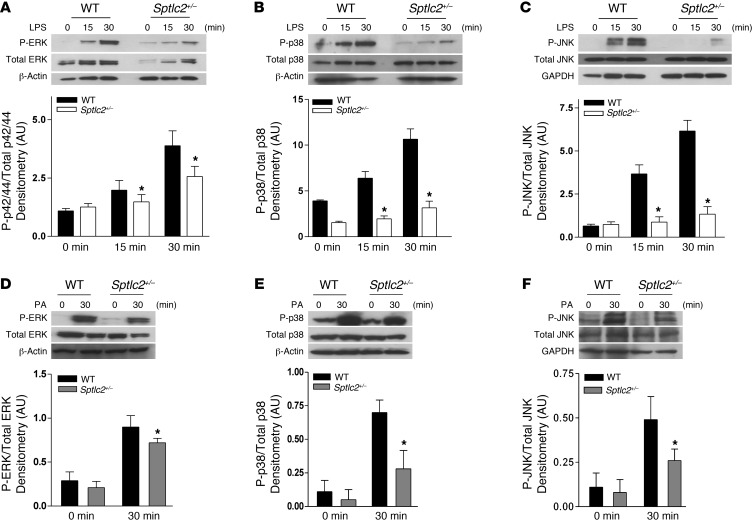
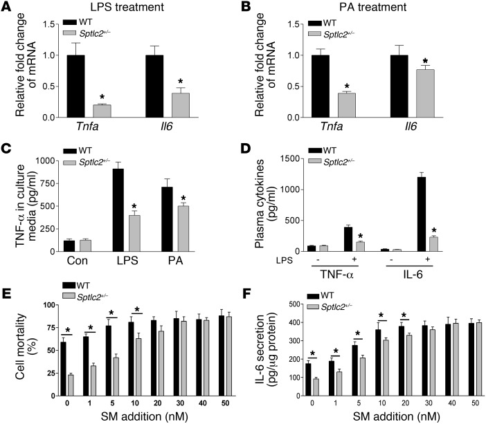
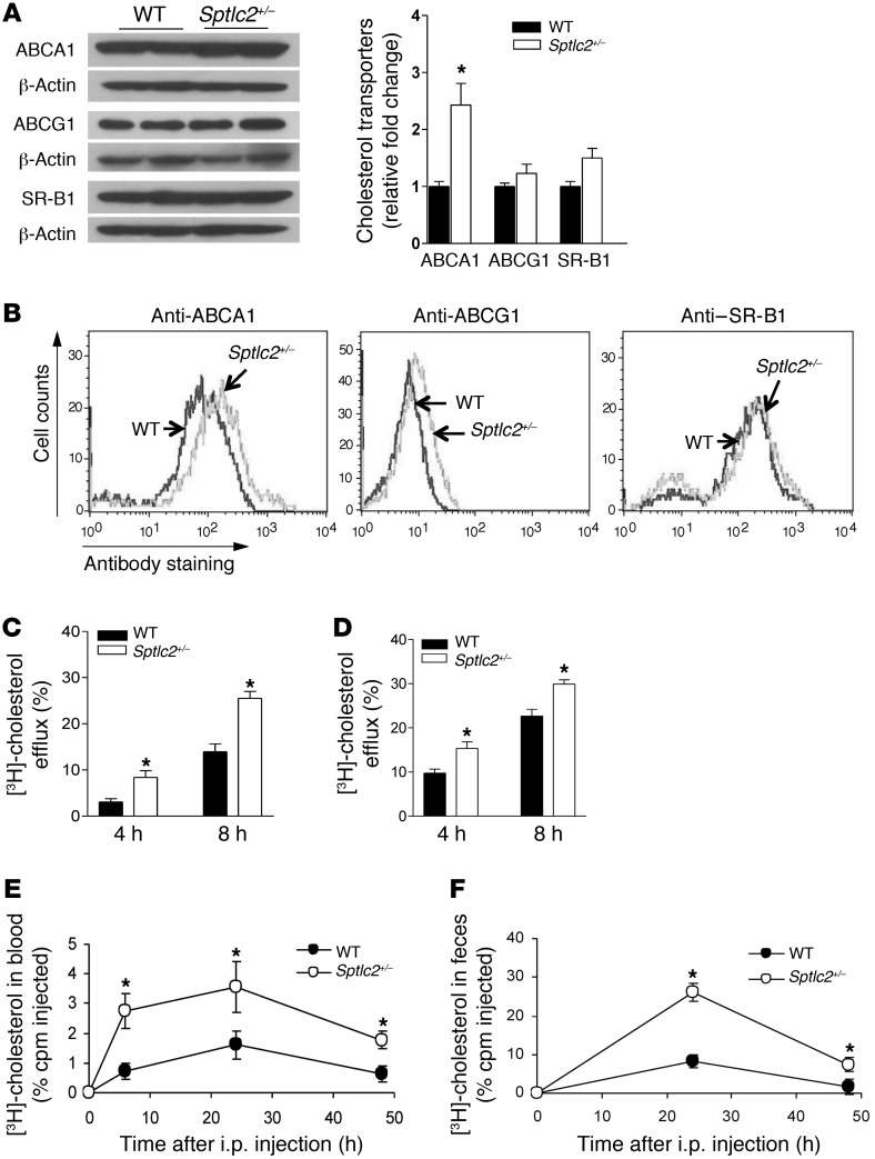
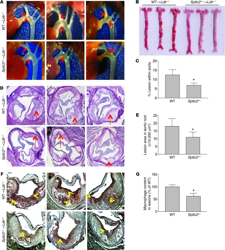
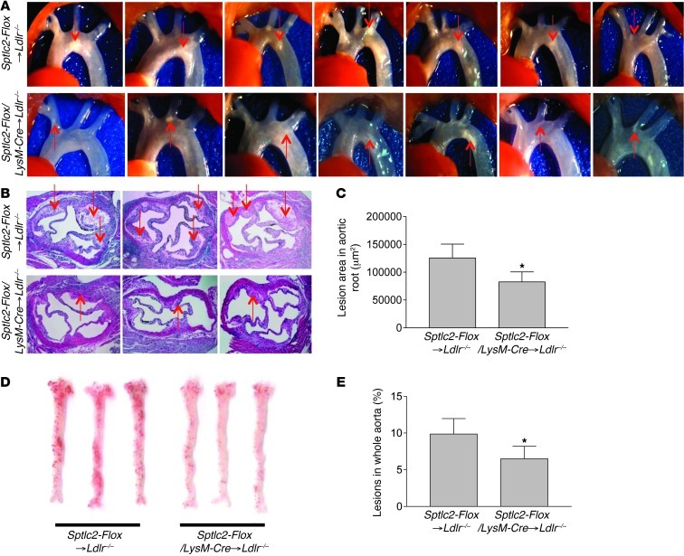
Serine palmitoyltransferase (SPT) is the first and rate-limiting enzyme of the de novo biosynthetic pathway of sphingomyelin (SM). Both SPT and SM have been implicated in the pathogenesis of atherosclerosis, the development of which is driven by macrophages; however, the role of SPT in macrophage-mediated atherogenesis is unknown. To address this issue, we have analyzed macrophage inflammatory responses and reverse cholesterol transport, 2 key mediators of atherogenesis, in SPT subunit 2-haploinsufficient (Sptlc2(+/-)) macrophages. We found that Sptlc2(+/-) macrophages have significantly lower SM levels in plasma membrane and lipid rafts. This reduction not only impaired inflammatory responses triggered by TLR4 and its downstream NF-κB and MAPK pathways, but also enhanced reverse cholesterol transport mediated by ABC transporters. LDL receptor-deficient (Ldlr(-/-)) mice transplanted with Sptlc2(+/-) bone marrow cells exhibited significantly fewer atherosclerotic lesions after high-fat and high-cholesterol diet feeding. Additionally, Ldlr(-/-) mice with myeloid cell-specific Sptlc2 haploinsufficiency exhibited significantly less atherosclerosis than controls. These findings suggest that SPT could be a novel therapeutic target in atherosclerosis.


Figures

Figure 1
Figure 1. Characterization of bone marrow–derived Sptlc2+/– macrophages.
Bone marrow–derived macrophages were isolated from WT and Sptlc2+/– mice. (A and B) Sptlc2+/– macrophages exhibited reduced expression of SPTLC2 and SPTLC1 subunits, as measured by Western blot from whole cell lysates with densitometric quantification. (C) Total RNA was extracted using TRIzol method, and Sptlc1, Sptlc2, and Sptlc3 mRNA were measured by real-time PCR using specific primers. (D) SPT activity in Sptlc2+/– macrophages with densitometric quantification. SPT activity in bone marrow–derived macrophage homogenate was measured using [14C]-serine and palmitoyl-CoA as substrates. The product, [14C]-keto-dihydrosphingosine (KDS), was separated from the reaction mix by thin layer chromatography, and the plate was scanned using Phosphorimager. (E) Intracellular SM content in macrophages was measured by LC/MS/MS. Data are representative of 3 independent experiments (n = 4 per group). *P < 0.05.
Figure 2
Figure 2. Sptlc2 haploinsufficiency in macrophages leads to significant reductions of SM, glucosylceramide, and GM3 in macrophage plasma membranes and lipid rafts, resulting in altered raft distribution.
(A) Cell surface SM measurement by lysenin-mediated cell lysis assay, presented as percent cell mortality. (B) Plasma membrane isolation from primary macrophages. The purity of cytoplasm (Cyto), total subcellular organelle membranes (TM), and plasma membranes (PM) was determined by Western blot; Na+/K+ATPase was used as a plasma membrane marker, and cytochrome C as a mitochondrial marker. (C) SM and phosphatidylcholine (PC) measurements in macrophage plasma membranes and lipid rafts using LC/MS/MS. (D) Ceramide (Cer), glucosylceramide (GlcCer), and GM3 in macrophage plasma membranes and lipid rafts, as measured by LC/MS/MS. Plasma membrane measurements were done using equal numbers of macrophages (~50 × 106 cells per group), and lipid raft measurements were performed using equal quantities of proteins from macrophage lysates. (E) Lipid rafts were isolated from macrophages using sucrose density gradient centrifugation, and fractions were collected. Western blots were performed for CAV-1 (raft marker) and CD71 (non-raft marker) with respective antibodies. Data are representative of 3 independent experiments (n = 6 per group). *P < 0.05.
Figure 3
Figure 3. Sptlc2+/– macrophages attenuate TLR4 recruitment and NF-κB activation after LPS stimulation.
(A and B) TLR4 receptors, analyzed by flow cytometry, on the surface of macrophages treated with 10 ng/ml LPS for 16 hours. (C) Nuclear NF-κB (p65) Western blot and quantitation. (D) Cytosolic IκBα Western blot and quantitation. (E and F) Immunocytochemistry (E) and quantitation (F) of NF-κB (p65) nuclear translocation. Data are representative of 3 independent experiments (n = 3 per group). *P < 0.05.
Figure 4
Figure 4. Sptlc2+/– macrophages attenuate MAPK activation after LPS or palmitate stimulation.
(A and D) ERK, (B and E) p38, and (C and F) JNK (phosphorylated and total protein) Western blot and quantitation after treatment with (AC) 1 μg/ml LPS or (DF) 500 μM palmitate (PA) in 1% BSA for 30 minutes (0-minute controls for palmitate were treated with 1% BSA only). GAPDH and β-actin were used for loading controls, as indicated. Data are representative of 3 independent experiments (n = 3 per group). *P < 0.05.
Figure 5
Figure 5. Sptlc2+/– macrophages exhibit reduced migration under inflammatory conditions.
(A) MCP-1 secreted by macrophages in culture medium after treatment with 10 ng/ml LPS for 16 hours. (B and C) Mouse plasma MCP-1 levels (B) under basal conditions and (C) after 8 hours of LPS challenge (50 μg/kg body weight i.p.). (D) In vitro Transwell migration of WT and Sptlc2+/– macrophages in response to LPS-treated macrophage culture medium, as seen by DAPI staining. (E) Quantitation of migrated cells in an average of 10 random microscopic fields. Data are representative of 3 independent experiments (n = 3 per group). For in vivo studies, n = 7. *P < 0.05.
Figure 6
Figure 6. Sptlc2+/– macrophages attenuate LPS- or palmitate-mediated expression of proinflammatory genes downstream of NF-κB and MAPKs.
(A and B) Real-time PCR for Tnfa and Il6 mRNA after treatment with (A) 1 μg/ml LPS for 15 minutes or (B) 500 μM palmitate for 30 minutes. (C) TNF-α secreted by macrophages in culture medium after treatment with 10 ng/ml LPS or 50 μM palmitate for 16 hours. (D) Mouse plasma TNF-α and IL-6 concentration after 8 hours of LPS challenge (50 μg/kg body weight i.p.). (E) Effect of exogenous SM on lysenin-mediated cell mortality. (F) Effect of exogenous SM on macrophage IL-6 secretion in response to treatment with 10 ng/ml LPS for 16 hours. Extracellular culture medium IL-6 was measured by ELISA. Data are representative of 3 independent experiments (n = 3). For in vivo studies, n = 7. *P < 0.05.
Figure 7
Figure 7. Sptlc2 haploinsufficiency increases macrophage cholesterol efflux, ex vivo and in vivo.
(A) Macrophage ABCA1, ABCG1, and SR-B1 Western blot and quantitation after treatment with 50 μg/ml acetylated-LDL for 16 hours. (B) Macrophage surface expression of ABCA1, ABCG1, and SR-B1 after treatment with 50 μg/ml acetylated-LDL for 16 hours, as measured by flow cytometry. (C and D) In vitro cholesterol efflux mediated by apoA-I (C) and HDL (D) in WT and Sptlc2+/– macrophages. (E and F) Sptlc2+/– or WT bone marrow–derived macrophages were loaded with cholesterol by incubation with acetylated-LDL and [3H]-cholesterol. Labeled macrophages were injected i.p. into C57BL/6 acceptor mice. (E) Plasma samples were collected at 6, 24, and 48 hours, and (F) feces were collected at 24 and 48 hours; all were analyzed for tracer counts using liquid scintillation counter. Data are representative of 3 independent experiments (n = 3 per group). For in vivo studies, n = 7. *P < 0.01.
Figure 8
Figure 8. Reduced atherosclerotic lesions in Sptlc2+/–Ldlr–/– mice.
(A) Aortic arches with atherosclerotic plaques (red arrows). (B) En face aortic plaque analysis after Oil Red O staining. (C) Quantitation of en face assay. (D) Aortic root assay for lesion areas after H&E staining. Red arrows denote atherosclerotic plaques. (E) Quantitation of root assay. (F) Immunohistochemical staining of macrophage accumulation in lesions (brown-stained regions, denoted by yellow arrows). (G) Quantitation of macrophage content. Quantitative analyses were done using ImageJ; 6 alternate sections (10 μm thick) sliced from paraffin-fixed aortic root tissues of each transplanted mouse were used for analysis. n = 9. *P < 0.05.
Figure 9
Figure 9. Reduced atherosclerotic lesions in Sptlc2-Flox/LysM-Cre→Ldlr–/– mice.
(A) Aortic arches with atherosclerotic plaques (red arrows). (B) Aortic root assay for lesion areas after H&E staining. 6 alternate sections (10 μm thick) sliced from paraffin-fixed aortic root tissues of each transplanted mouse were used for analysis. Red arrows denote atherosclerotic plaques. (C) Quantitation of root assay. (D) En face aortic plaque analysis after Oil Red O staining. (E) Quantitation of en face assay. n = 9. *P < 0.05.

References

    1. Libby P. Inflammation in atherosclerosis. Nature. 2002;420(6917):868–874. doi: 10.1038/nature01323. - DOI - PubMed
    1. Mayo MW, Baldwin AS. The transcription factor NF-kappaB: control of oncogenesis and cancer therapy resistance. Biochim Biophys Acta. 2000;1470(2):M55–M62. - PubMed
    1. Karin M, Lin A. NF-kappaB at the crossroads of life and death. Nat Immunol. 2002;3(3):221–227. doi: 10.1038/ni0302-221. - DOI - PubMed
    1. Grilli M, Chiu JJ, Lenardo MJ. NF-kappa B and Rel: participants in a multiform transcriptional regulatory system. Int Rev Cytol. 1993;143:1–62. - PubMed
    1. Verma IM, Stevenson JK, Schwarz EM, Van Antwerp D, Miyamoto S. Rel/NF-kappa B/I kappa B family: intimate tales of association and dissociation. Genes Dev. 1995;9(22):2723–2735. doi: 10.1101/gad.9.22.2723. - DOI - PubMed

Publication types

MeSH terms