Review
. 2013 Apr 2;14(4):7231-72.
doi: 10.3390/ijms14047231.

Peripheral reproductive organ health and melatonin: ready for prime time

Affiliations
Review

Peripheral reproductive organ health and melatonin: ready for prime time

Russel J Reiter et al. Int J Mol Sci. .

Abstract

Melatonin has a wide variety of beneficial actions at the level of the gonads and their adnexa. Some actions are mediated via its classic membrane melatonin receptors while others seem to be receptor-independent. This review summarizes many of the published reports which confirm that melatonin, which is produced in the ovary, aids in advancing follicular maturation and preserving the integrity of the ovum prior to and at the time of ovulation. Likewise, when ova are collected for in vitro fertilization-embryo transfer, treating them with melatonin improves implantation and pregnancy rates. Melatonin synthesis as well as its receptors have also been identified in the placenta. In this organ, melatonin seems to be of particular importance for the maintenance of the optimal turnover of cells in the villous trophoblast via its ability to regulate apoptosis. For male gametes, melatonin has also proven useful in protecting them from oxidative damage and preserving their viability. Incubation of ejaculated animal sperm improves their motility and prolongs their viability. For human sperm as well, melatonin is also a valuable agent for protecting them from free radical damage. In general, the direct actions of melatonin on the gonads and adnexa of mammals indicate it is an important agent for maintaining optimal reproductive physiology.


Figures

Figure 1
Figure 1
A three-dimensional view of melatonin (N-acetyl-5-methoxytryptamine), an indoleamine originally discovered to be a secretory product of the mammalian pineal gland and subsequently found to be produced in many different cells/organs and in all species of the plant and animal kingdoms. Melatonin easily crosses cell membranes and all morphophysiological barriers, e.g., the blood-brain barrier. While the current review summarizes its actions at the level of both the female and male reproductive system, the beneficial functions of this molecule probably extend to every cell in the organism.
Figure 2
Figure 2
This figure illustrates the actions of melatonin in reducing free radical-mediated molecular damage. Melatonin stimulates (blue lines) several antioxidative enzymes including superoxide dismutase (SOD), glutathione peroxidase (GPx), glutathione reductase (GRd) and glutamylglycine ligase (GCL). It also inhibits (red line) the pro-oxidative enzyme nitric oxide synthase (NOS). In addition to modulating the activity of these enzymes, melatonin directly scavenges the highly toxic hydroxyl radical (·OH), the peroxynitrite anion (ONOO) and possibly some other radical and non-radical products. The superoxide anion radical (O2•−), hydrogen peroxide (H2O2) and the ·OH are referred to as reactive oxygen species (ROS); nitric oxide (NO) and ONOO are referred to as reactive nitrogen species (RNS). O2 = molecular oxygen; e = electron; Fe2+ = ferrous iron.
Figure 3
Figure 3
Schematic representation of the synthesis of melatonin from the amino acid, tryptophan. Tryptophan, which is taken up from the blood, via the four step pathway outline is converted to N-acetyl-5-methoxytryptamine (melatonin). Melatonin is best known for its production in the cells of the pineal gland from which it is quickly released in body fluids, i.e., blood and cerebrospinal fluid. Circulating melatonin has both receptor-mediated and receptor-independent actions. Many other cells also produce melatonin; in this case, the indoleamine does not gain access to the blood in any appreciable amounts but rather works near its site of synthesis as an autacoid or as a paracoid.
Figure 4
Figure 4
In the ovarian follicle, melatonin (represented in this figure by the M) impacts the function of numerous cells, especially granulosa cells and the ovum (oocyte). The actions of melatonin in these cells are mediated via membrane receptors (MT, in particular MT1 and MT2) and also possibly via binding sites in the nucleus and in the cytosol. In addition to its receptor-mediated actions, melatonin also functions as a direct free radical scavenger to reduce oxidative stress at the level of the ovary; this beneficial action is carried out without an interaction with a receptor. Additional antioxidant functions of melatonin are achieved when the indole stimulates enzymes which metabolize free radicals to less toxic products. The antioxidative enzymes include superoxide dismutase (SOD), glutathione peroxidase (GPx) and catalase (CAT) in thecal cells, granulosa cells and in the follicular fluid. Via these actions, melatonin reduces free radical damage, which would be especially bad for the ovum, and maintains these elements in an optimally functional state. The origin of melatonin in the follicular fluid is the blood and from its local synthesis in granulosa cells. C, cholesterol; LH R, LH receptor; FSH R, FSH receptor; NAT, N-acetyltransferase; HIOMT, hydroxyindole-O-methyltransferase (currently known as acetylserotonin methyltransferase, ASMT); MIH, maturation-inducing hormone; MPF, maturation-promoting factor; GVBD, germinal vesicle breakdown; ROS, reactive oxygen species; IGF, insulin-like growth factor; TGF-β, transforming growth factor β. From Tamura et al.[80].
Figure 5
Figure 5
Mean (±SEM) 8-OHdG (8-hydroxy-2-deoxyguanosine; a product of oxidatively damaged DNA) and HEL (hexanoyl-lysine adduct; a product of oxidatively damaged lipid) levels in the follicular fluid of women treated with melatonin (3 mg daily) or vitamin E (600 mg daily) for 37 days. Both antioxidants obviously reduced free radical damage; however, even though the dose of melatonin was 200 times less than that of vitamin E, it proved as effective in reducing free radical damage to the oocytes. * p < 0.05 compared to control values. Modified from Tamura et al.[96].
Figure 6
Figure 6
The in vitro exposure of denuded mouse oocytes to the oxidizing agent, H2O2 (300 μM), caused a marked drop (from 92.3% to 28.6% in the percentage of cells that matured (presence of first polar body; MII stage oocytes) within 12 h. Increasing concentrations of melatonin in the medium clearly improved the percent of H2O2-exposed oocytes that reached maturity. The number of oocytes in each treatment group varied from 36 to 42. Different letters indicate statistically significant differences between groups (p < 0.01; Chi-square test). Modified from Tamura et al.[96].
Figure 7
Figure 7
Distribution of mitochondria (identified with Mito Tracker® Red) in MII bovine oocytes matured in the absence (A) or presence (B) of melatonin in the culture medium. The medium was supplemental with melatonin at a concentration of 10 ng/mL. The subcellular distribution of the mitochondria was markedly impacted by the presence of melatonin, probably due to the actions of the indoleamine or the cytoskeleton. In the control cells, the mitochondria were clustered around the periphery of the ovum whereas melatonin treatment caused their dispersion throughout the cell. The arrowheads identify the first polar body. From El-Raey et al.[97] with permission.
Figure 8
Figure 8
Turnover of cells that make up the villous trophoblast is essential for proper functioning of the placenta. Proliferative cytotrophoblast stem cells differentiate and eventually exit the cell cycle. These cells then fuse to form a multinucleated syncytium, the syncytiotrophoblast. Within several days cells of the syncytiotrophoblast undergo apoptosis. The additions of differentiated cytotrophoblasts replace the syncytiotrophoblast cells that are lost via apoptosis. The apoptotic loss of the syncytiotrophoblast is balanced by the fusion of differentiated cytotrophoblast cells. Locally produced melatonin seems to be involved in maintenance of homeostasis by limiting apoptosis of the differentiated cytotrophoblasts while enhancing apoptosis of the syncytiotrophoblast; these latter cells have characteristics of cancer cells in which melatonin also causes apoptosis. From Lanoix et al.[135].
Figure 9
Figure 9
Diagrammatic representation of the circadian melatonin rhythm in human females during the last trimester of pregnancy and after delivery. The nocturnal melatonin peak gradually increases near term pregnancy. The augmented melatonin levels seem to aid in inducing uterine contractions at parturition since it has been shown to synergize with the released oxytocin to cause stronger uterine contractions. Shortly after delivery of the fetus, the nocturnal melatonin peak returns to its pre-pregnancy levels.
Figure 10
Figure 10
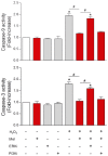
Evidence that the melatonin membrane receptor, MT1, is involved in the protective actions against H2O2 toxicity in human spermatozoa. Both the activity of caspase 9 (top panel) and caspase 3 (bottom panel), changes that are indicative of pending apoptosis, were elevated when the sperm were exposed to H2O2. These increases were blocked when luzindole (Luz), an MT1 and MT2 receptor antagonist, was added to the incubation medium but not when the selective MT2 blocker, 4P-PDOT, was added. These findings are consistent with the MT1 receptor mediating, at least in part, the ability of melatonin to defer H2O2-induced apoptotic processes in human spermatozoa. *p < 0.05, compared with all other values; # p < 0.05. From Espino et al.[225].
Figure 11
Figure 11
To define the signal transduction pathway relative to melatonin’s actions on human spermatozoa, sperm were exposed to H2O2 in the presence of either PD98059, an ERK inhibitor, or to LY294002, a selective pharmacological inhibitor of the P13K/Akt pathway. Clearly, suppressing ERK interfered with the H2O2-mediated rise in caspase 9 (top panel) and caspase 3 (bottom panel) activity indicating that this pathway is related to the ability of melatonin to forestall apoptotic processes in human spermatozoa. *p < 0.05 compared to all other values; # p < 0.05. From Espino et al.[225].
Figure 12
Figure 12
Correlations between urinary 6-hydroxymelatonin sulfate (aMT6-s), a major hepatic metabolite of melatonin, in the first urine morning void and various human sperm parameters in 20 adult males (20–40 years age). The semen samples were collected by masturbation after 4–5 days of sexual abstinence. The following parameters correlated positively with the concentration of urinary aMT6-s, which was taken as an index of endogenous melatonin levels: sperm concentration (A), sperm motility (B), sperm morphology (C), sperm vitality (D), and the total antioxidant status of the urine (estimated using the ABTS assay). There was a negative correlation between urinary aMT6-s and the number of round spermatids (cells) in the semen (E). Data from Espino et al.[225].

References

    1. Lerner A.B., Case J.D., Takahashi Y., Lee Y., Mori W. Isolation of melatonin, the pineal factor that lightens melanocytes. J. Am. Chem. Soc. 1958;80:2587.
    1. Launay J.M., Lemaitre B.J., Husson H.P., Dreux C., Hartmann L., da Prada M. Melatonin synthesis by rabbit platelets. Life Sci. 1982;31:1487–1494. - PubMed
    1. Reiter R.J., Richardson B.A., Matthews S.A., Lane S.J., Ferguson B.N. Rhythms of immunoreactive melatonin in the retina and Harderian glands of rats: Persistence after pinealectomy. Life Sci. 1983;32:1299–1236. - PubMed
    1. Huether G., Poeggeler B., Reimer A., George A. Effects of tryptophan administration on circulating melatonin levels in chicks and rats: Evidence for stimulation of melatonin synthesis and release in the gastrointestinal tract. Life Sci. 1992;51:946–953. - PubMed
    1. Abe M., Itoh M.T., Miyata M., Shimizu K., Sumi Y. Circadian rhythm of serotonin N-acetyltransferace activity in rat lens. Exp. Eye Res. 2000;70:805–808. - PubMed

LinkOut - more resources