

The role of PET/CT in cervical cancer
- PMID: 23549376
- PMCID: PMC3581867
- DOI: 10.3389/fonc.2013.00034
The role of PET/CT in cervical cancer
Abstract
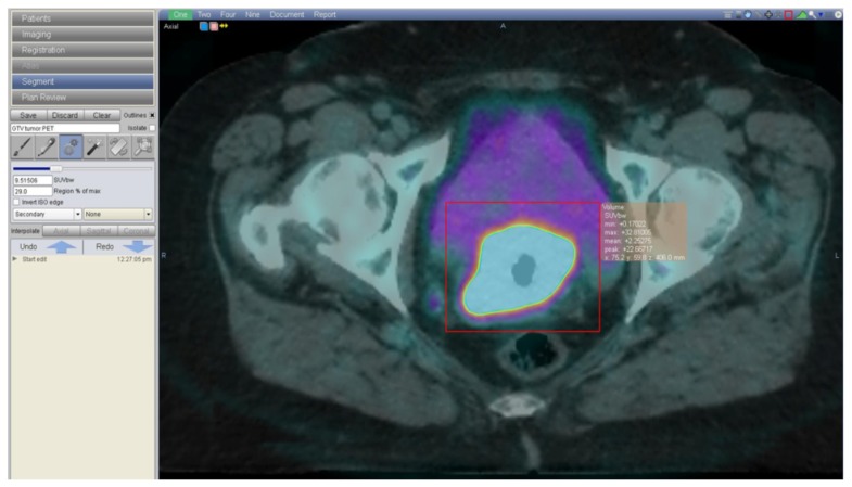
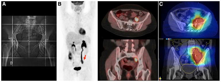
In locally advanced cervical cancer, (18)F-fluorodeoxyglucose (FDG) positron emission tomography - computed tomography (PET/CT) has become important in the initial evaluation of disease extent. It is superior to other imaging modalities for lymph node status and distant metastasis. PET-defined cervical tumor volume predicts progression-free and overall survival. Higher FDG uptake in both primary and regional lymph nodes is strongly predictive of worse outcome. FDG-PET is useful for assessing treatment response 3 months after completing concurrent chemo-radiotherapy (CRT) and predicting long-term survival, and in suspected disease recurrence. In the era of image-guided adaptive radiotherapy, accurately defining disease areas is critical to avoid irradiating normal tissue. Based on additional information provided by FDG-PET, radiation treatment volumes can be modified and higher doses to FDG-positive lymph nodes safely delivered. FDG-PET/CT has been used for image-guided brachytherapy of FDG-avid tumor volume, while respecting low doses to bladder and rectum. Despite survival improvements due to CRT in cervical cancer, disease recurrences continue to be a major problem. Biological rationale exists for combining novel non-cytotoxic agents with CRT, and drugs targeting specific molecular pathways are under clinical development. The integration of these targeted therapies in clinical trials, and the need for accurate predictors of radio-curability is essential. New molecular imaging tracers may help identifying more aggressive tumors. (64)Cu-labeled diacetyl-di(N(4)-methylthiosemicarbazone) is taken up by hypoxic tissues, which may be valuable for prognostication and radiation treatment planning. PET/CT imaging with novel radiopharmaceuticals could further impact cervical cancer treatment as surrogate markers of drug activity at the tumor microenvironment level. The present article reviews the current and emerging role of PET/CT in the management of cervical cancer.
Keywords: Fluorodeoxyglucose F18; Planning Treatment Volumes; cervical cancer; positron emission tomography; radiation therapy.
Figures


References
-
- Alva A., Slovin S., Daignault S., Carducci M., Dipaola R., Pienta K., et al. (2012). Phase II study of cilengitide (EMD 121974, NSC 707544) in patients with non-metastatic castration resistant prostate cancer, NCI-6735. A study by the DOD/PCF prostate cancer clinical trials consortium. Invest. New Drugs 30 749–757 - PMC - PubMed
-
- Bachtiary B., Dewitt A., Pintilie M., Jezioranski J., Ahonen S., Levin W., et al. (2005). Comparison of late toxicity between continuous low-dose-rate and pulsed-dose-rate brachytherapy in cervical cancer patients. Int. J. Radiat. Oncol. Biol. Phys. 63 1077–1082 - PubMed
-
- Beekman K. W., Colevas A. D., Cooney K., Dipaola R., Dunn R. L., Gross M., et al. (2006). Phase II evaluations of cilengitide in asymptomatic patients with androgen-independent prostate cancer: scientific rationale and study design. Clin. Genitourin. Cancer 4 299–302 - PubMed
-
- Blower P. J., Lewis J. S., Zweit J. (1996). Copper radionuclides and radiopharmaceuticals in nuclear medicine. Nucl. Med. Biol. 23 957–980 - PubMed
LinkOut - more resources
Full Text Sources
Other Literature Sources
Research Materials