Dysplastic nevi and melanoma
- PMID: 23549396
- PMCID: PMC3616416
- DOI: 10.1158/1055-9965.EPI-12-1346
Dysplastic nevi and melanoma
Abstract
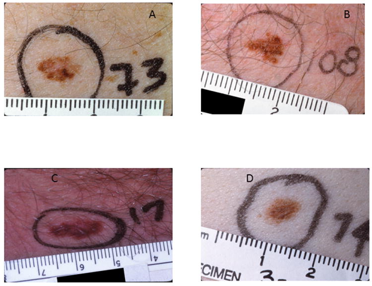
Dysplastic nevi are described as being on a continuum between common acquired nevi and melanoma because they are morphologically and biologically intermediate between these 2 entities. Since initially being reported as histologic lesions observed in melanoma-prone families, there has been considerable debate about the definition of dysplastic nevi, the histologic and clinical criteria used to define them, and their biologic importance. Their role as precursor lesions for melanoma is not their primary role in their relationship to melanoma because of the rarity of transformation of any individual nevus to a melanoma. Although there is still no single, universally agreed upon histologic or clinical definition or even name for these nevi, dysplastic nevi should be considered important because of their association with an increased risk for melanoma.
Conflict of interest statement
The authors declare that they have no conflicts of interest related to this manuscript.
Figures
References
-
- Clark WH, Jr, Reimer RR, Greene M, Ainsworth AM, Mastrangelo MJ. Origin of familial malignant melanomas from heritable melanocytic lesions. ‘The B-K mole syndrome’. Archives of dermatology. 1978;114:732–8. - PubMed
-
- Frichot BC, 3rd, Lynch HT, Guirgis HA, Harris RE, Lynch JF. New cutaneous phenotype in familial malignant melanoma. Lancet. 1977;1:864–5. - PubMed
-
- Elder DE. Dysplastic naevi: an update. Histopathology. 2010;56:112–20. - PubMed
-
- Elder DE, Goldman LI, Goldman SC, Greene MH, Clark WH., Jr Dysplastic nevus syndrome: a phenotypic association of sporadic cutaneous melanoma. Cancer. 1980;46:1787–94. - PubMed
Publication types
MeSH terms
Grants and funding
LinkOut - more resources
Full Text Sources
Other Literature Sources
Medical