Review
. 2013 Jul;41(7):1347-65.
doi: 10.1007/s10439-013-0800-z. Epub 2013 Apr 3.

Physical factors effecting cerebral aneurysm pathophysiology

Affiliations
Review

Physical factors effecting cerebral aneurysm pathophysiology

Chander Sadasivan et al. Ann Biomed Eng. 2013 Jul.

Abstract

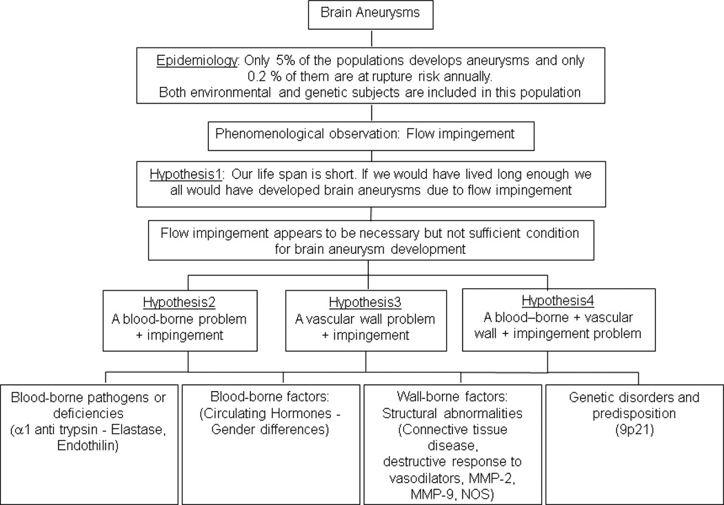
Many factors that are either blood-, wall-, or hemodynamics-borne have been associated with the initiation, growth, and rupture of intracranial aneurysms. The distribution of cerebral aneurysms around the bifurcations of the circle of Willis has provided the impetus for numerous studies trying to link hemodynamic factors (flow impingement, pressure, and/or wall shear stress) to aneurysm pathophysiology. The focus of this review is to provide a broad overview of such hemodynamic associations as well as the subsumed aspects of vascular anatomy and wall structure. Hemodynamic factors seem to be correlated to the distribution of aneurysms on the intracranial arterial tree and complex, slow flow patterns seem to be associated with aneurysm growth and rupture. However, both the prevalence of aneurysms in the general population and the incidence of ruptures in the aneurysm population are extremely low. This suggests that hemodynamic factors and purely mechanical explanations by themselves may serve as necessary, but never as necessary and sufficient conditions of this disease's causation. The ultimate cause is not yet known, but it is likely an additive or multiplicative effect of a handful of biochemical and biomechanical factors.


Figures

Figure 1
Figure 1
Circle of Willis showing common locations of cerebral aneurysms. From Schievink 1997(N. Engl. J. Med., Schievink WI, Intracranial aneurysms, 336, pp.29, Copyright ©1997 Massachusetts Medical Society. Reprinted with permission from Massachusetts Medical Society)
Figure 2
Figure 2
A basic flowchart showing mechanistic possibilities of aneurysm pathophysiology
Figure 3
Figure 3
Schematics of flow patterns in A) sidewall, B) bifurcation with symmetrical outflow, C) bifurcation with asymmetrical outflow, and D) asymmetric bifurcation. E) Laser induced fluorescence study in a sidewall channel at mid systolic acceleration; F) Dye visualization in an elastomer model of an anterior communicating artery aneurysm; asymmetrical internal carotid flow; G) Computational fluid dynamics in a giant internal carotid-posterior communicating artery aneurysm at early diastole; H) schematic of flow patterns in a high aspect ratio bifurcation aneurysm with a bleb; I) schematic at one instant in the cardiac cycle of different flow patterns obtained from computational fluid dynamics simulations in anatomically realistic aneurysms. Panels A–D from Steiger 1987 (With kind permission from Springer Science+Business Media: Heart Vessels, Basic flow structure in saccular aneurysms, 3, 1987, pp.57–61, Steiger HJ et al., Figures 3,7a,7b,8); panel E from Lieber 1997 (With kind permission from Springer Science+Business Media: Ann. Biomed. Eng., Alteration of hemodynamics in aneurysm models by stenting, 25, 1997, pp.463, Lieber BB et al., Figure 5A); panel F from Kerber 1999 (CW Kerber et al., Flow dynamics in a lethal anterior communicating artery aneurysm, AJNR Am J Neuroradiol, 20, 10, pp.2000–3, 1999 © by American Society of Neuroradiology); panel G from Steinman 2003 (DA Steinman et al., Image-based computational simulation of flow dynamics in a giant intracranial aneurysm, AJNR Am J Neuroradiol, 24, 4, pp.559–66, 2003 © by American Society of Neuroradiology); panel H from Ujiie 1999 (Ujiie H et al., Effects of size and shape (aspect ratio) on the hemodynamics of saccular aneurysms, Neurosurgery, 45, 1, pp.119-29, 1999); panel I from Cebral 2005 (JR Cebral et al., Characterization of cerebral aneurysms for assessing risk of rupture by using patient-specific computational hemodynamics models, AJNR Am J Neuroradiol, 26, 10, pp.2550–9, 2005 © by American Society of Neuroradiology)
Figure 4
Figure 4
A) Velocity vectors obtained with laser Doppler velocimetry in acrylic casts of a middle cerebral artery aneurysm before and after it grew over a period of 1 year. Examples of wall shear stress magnitudes calculated with computational fluid dynamics in B) a posterior communicating artery and C) a middle cerebral artery aneurysm. Panel A from Tateshima 2007 (S Tateshima et al., Intra-aneurysmal hemodynamics during the growth of an unruptured aneurysm, AJNR Am J Neuroradiol, 28, 4, pp.622–7, 2007 © by American Society of Neuroradiology); Panel B from Kulcsár 2011 (Z Kulcsár, et al., I Szikora, Hemodynamics of cerebral aneurysm initiation, AJNR Am J Neuroradiol, 32, 3, pp.587–94, 2011 © by American Society of Neuroradiology); Panel C from Shojima 2004 (Shojima M et al., Magnitude and role of wall shear stress on cerebral aneurysm, Stroke, 35, 11, pp.2500–5, 2004)
Figure 5
Figure 5
Four different types of aneurysm wall generally characterizing changes during progression to rupture. AN: aneurysm; PA: parent artery; N: neck; D: dome; ad: adventitial side; lu: luminal side; mh: myointimal hyperplasia; t: fresh thrombus; ot: organizing thrombus Left column with types I–IV from Suzuki 1978 (Suzuki J and Ohara H, Clinicopathological study of cerebral aneurysms, J. Neurosurg., 48, pp.505–14, 1978); right columns with types A–D from Tulamo 2010 (Adapted by permission from BMJ Publishing Group Limited. [J. Neurointerv. Surg., Tulamo R, Frösen J, Hernesniemi J, Niemelä M, 2, pp.120–30, 2010])

References

    1. Acar F, Men S, Tayfur V, Yilmaz O, Erbayraktar S, Metin Güner E. In vivo intraaneurysmal pressure measurements in experimental lateral wall aneurysms before and after onyx embolization. Surg Neurol. 2006;66:252–256. - PubMed
    1. Alnaes MS, Isaksen J, Mardal KA, Romner B, Morgan MK, Ingebrigtsen T. Computation of hemodynamics in the circle of Willis. Stroke. 2007;38:2500–2505. - PubMed
    1. Antiga L, Piccinelli M, Botti L, Ene-Iordache B, Remuzzi A, Steinman DA. An image-based modeling framework for patient-specific computational hemodynamics. Medical & Biological Engineering & Computing. 2008;46:1097–1112. - PubMed
    1. Baharoglu MI, Lauric A, Gao BL, Malek AM. Identification of a dichotomy in morphological predictors of rupture status between sidewall- and bifurcation-type intracranial aneurysms. J Neurosurg. 2012;116:871–881. - PubMed
    1. Bai-Nan X, Fu-Yu W, Lei L, Xiao-Jun Z, Hai-Yue J. Hemodynamics model of fluid-solid interaction in internal carotid artery aneurysms. Neurosurg Rev. 2011;34:39–47. - PMC - PubMed

Publication types

LinkOut - more resources