. 2013 Jan;4(1):4-18.
doi: 10.1111/jdi.12042. Epub 2013 Jan 29.

Diabetic cardiac autonomic neuropathy, inflammation and cardiovascular disease

Affiliations

Diabetic cardiac autonomic neuropathy, inflammation and cardiovascular disease

Aaron I Vinik et al. J Diabetes Investig. 2013 Jan.

Abstract

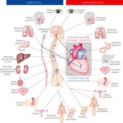
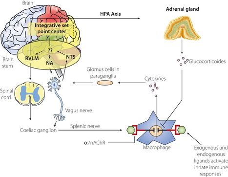
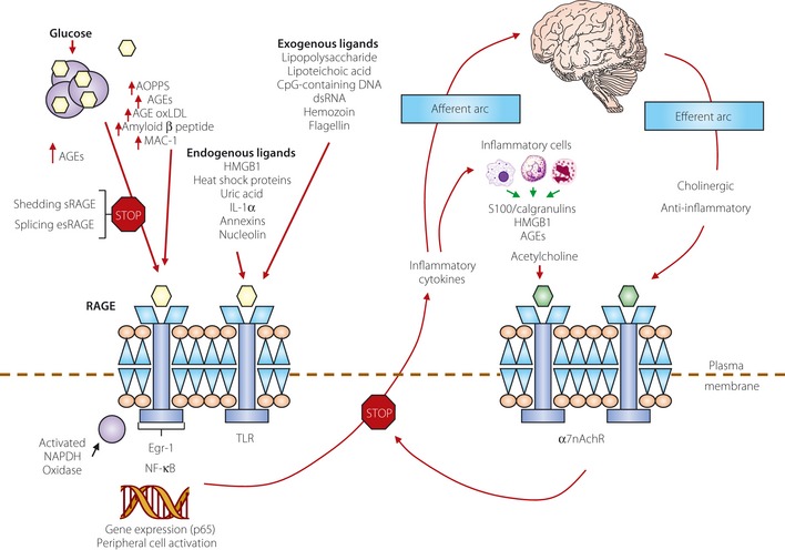
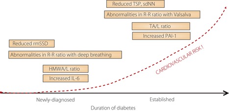
One of the most overlooked of all serious complications of diabetes is cardiovascular autonomic neuropathy. There is now clear evidence that suggests activation of inflammatory cytokines in diabetic patients and that these correlate with abnormalities in sympathovagal balance. Dysfunction of the autonomic system predicts cardiovascular risk and sudden death in patients with type 2 diabetes. It also occurs in prediabetes, providing opportunities for early intervention. Simple tests that can be carried out at the bedside with real-time output of information - within the scope of the practicing physician - facilitate diagnosis and allow the application of sound strategies for management. The window of opportunity for aggressive control of all the traditional risk factors for cardiovascular events or sudden death with intensification of therapy is with short duration diabetes, the absence of cardiovascular disease and a history of severe hypoglycemic events. To this list we can now add autonomic dysfunction and neuropathy, which have become the most powerful predictors of risk for mortality. It seems prudent that practitioners should be encouraged to become familiar with this information and apply risk stratification in clinical practice. Several agents have become available for the correction of functional defects in the autonomic nervous system, and restoration of autonomic balance is now possible.

Keywords: Cardiac autonomic neuropathy; Inflammation; Pathogenesis.


Figures

Figure 1
Figure 1
Physiological functions of both the sympathetic and parasympathetic nervous system.
Figure 2
Figure 2
Afferent sensory signals are transmitted by the vagus to the nucleus of the solitary tract (NTS) and polysynaptic receptors relay to the sympathetic through the rostral ventromedullary nucleus (RVLS), the parasympathetic nucleus ambiguous (NA) and the dorsal vagal nucleus. There is both sympathetic and parasympathetic output to the celiac ganglion. The splenic nerve activates the inflammatory cascade in macrophages, which might be in the spleen, but occur diffusely throughout the body. Stimulation of the vagus inhibits this activation by acetylcholine binding to the nicotinic acetylcholine receptor subunit a7 receptor, which curtails the response. Also shown is the co‐activation of the hypothalamic pituitary axis (HPA) with release of glucocorticoids, which also modulate the inflammatory response. It is unclear how this set point is determined in the body, but what is clear is that autonomic modulation of inflammation can be achieved by altering sympathetic parasympathetic balance. Adapted from Tracey6.
Figure 3
Figure 3
The relationship between binding of ligands to the pattern recognition advance glycation end‐product receptor (RAGE), and inflammation, gene expression, oxidative and nitrosative stress, and damage to the macro‐ and microvasculature. Elevated levels of glucose bind to proteins and form advance glycation end‐products (AGEs), which bind to RAGEs. RAGE signaling activates nicotinamide adenine dinucleotide phosphate‐oxidase and production of reactive oxygen species (ROS). Increased ROS increases advance oxidation protein products (AOPPs), more AGEs and AGE‐modification of oxidized low‐density lipoprotein (oxLDLs). Furthermore, increased ROS might deplete glutathione, thereby suppressing glyoxalase I activity, a mechanism favoring further AGE accumulation. AGEs, AOPPs, macrophage glycoprotein (MAC‐1) and AGE‐oxLDL ligands of RAGE sustain stimulation of RAGE, and these processes, together with increased ROS, activate key transcription factors, such as nuclear factor‐κB (NF‐κB) and Egr‐1, which increase gene transcription factors and activate inflammatory mechanisms. The consequences include increased migration and activation of RAGE‐expressing neutrophils, monocytes, macrophages, T‐cells and dendritic cells. This results in the release of the pro‐inflammatory RAGE ligands S100/calgranulins and high‐mobility group protein box‐1 (HMGB1). In this inflammatory environment, further AGEs might be formed as well. Through interaction with RAGE, these ligands magnify activation of NF‐κB, epidermal growth factor 1 (Egr‐1) and other factors, thereby amplifying cellular stress and tissue damage leading to neurovascular dysfunction. Soluble RAGE (sRAGE) formed from cleavage of RAGE by disintegrins, such as ADAM 10, a metalloproteinase, and β‐ and γ‐secretases. sRAGE or a spliced variant (esRAGE) compete for binding of ligands to RAGE, and a deficiency could theoretically initiate the sequence of events activating an inflammatory cascade with an increase in the expression of pro‐inflammatory cytokines (E‐selectin, endothelin‐1 tissue factor, vascular endothelial growth factor, interleukin‐6 and tumor necrosis factor‐α) and damage to neurons, kidney, eye, the vasculature, and even bone. Increasing sRAGE or its administration could competitively reduce activation of the AGE/RAGE pathway and it consequences. In addition, endogenous and exogenous ligands bind to Toll‐like receptors (TLR) also targeting NF‐κB, as well as inducing the secretin of pro‐inflammatory cytokines, which activate afferent sensory neurons reaching the brainstem through axons in the vagus. This in turn activates the cholinergic anti‐inflammatory efferent arc, which inhibits response in cytokine‐producing immune cells and signals through the nicotinic acetylcholine receptor subunit α7 (α7nACHR). This in turn inhibits NF‐κB activation. Thus, the pathways of AGE/RAGE activation of the inflammatory cascade and the inflammatory ligands target activation of an inflammatory cascade that can be abrogated by either competing with the binding of ligands to RAGE or by vagal activation of the anti‐inflammatory reflex. Thus, central to curtailing unbridled activation of the inflammatory cascade is the integrity of parasympathetic autonomic function or balance between the two arms of the autonomic nervous system. Reproduced from Vinik65.
Figure 4
Figure 4
The natural history of autonomic balance. HMWA/L, high molecular weight adiponectin/leptin ratio; IL‐6, interleukin‐6; PAI‐1, plasminogen activator inhibitor 1; R‐R, beat‐to‐beat interval; rmSSD, root mean square of the difference of successive beat‐to‐beat interval; sdNN, standard deviation of all normal beat‐to‐beat intervals; TA/L, total adiponectin/leptin ratio. Reproduced from Lieb et al.18.
Figure 5
Figure 5
Prevalence rate ratios and 95% confidence intervals for the association between cardiac autonomic neuropathy (CAN) and silent myocardial ischemia (MI) in 12 studies. Modified from Vinik et al.3.
Figure 6
Figure 6
Stages of cardiovascular autonomic neuropathy (reproduced from Spallone et al.25).
Figure 7
Figure 7
Spectral analysis of heart rate variability. HRV, heart rate variability; PS, parasympathetic; S, sympathetic; sdNN, standard deviations of all normal beat‐to‐beat intervals (modified from Vinik et al.3).
Figure 8
Figure 8
The effects of walking with and without a pet dog on parasympathetic function (mean high frequency power). (a) The 80 min walking program including two 30 min walks without and with the dog (n = 13). (b) The 80 min walking program over three consecutive days (n = 3). (c) Interaction with the dog at home during 6 h of continuous monitoring (n = 4; reproduced from Motooka et al.59. © Copyright 2006. The Medical Journal of Australia ‐ reproduced with permission).
Figure 9
Figure 9
The ability to correct sympathetic excess with (a) a β‐adrenergic blocking agent, (b) sympathetic withdrawal with an adrenergic agonist and (c) parasympathetic excess with an anti‐cholinergic agent. BP, blood pressure; HR, heart rate; PE, parasympathetic excess; SE, sympathetic excess; SW, sympathetic withdrawal. Reproduced from Vinik et al.11.

References

    1. Vinik AI, Maser RE, Mitchell BD, et al Diabetic Autonomic Neuropathy. Diabetes Care 2003; 26: 1553–1579 - PubMed
    1. Maser RE, Mitchell BD, Vinik AI, et al The association between cardiovascular autonomic neuropathy and mortality in individuals with diabetes: a meta‐analysis. Diabetes Care 2003; 26: 1895–1901 - PubMed
    1. Vinik AI, Ziegler D. Diabetic cardiovascular autonomic neuropathy. Circulation 2007; 115: 387–397 - PubMed
    1. Saravia F, Homo‐Delarche F. Is innervation an early target in autoimmune diabetes? Trends Immunol 2003; 24: 574–579 - PubMed
    1. Borovikova LV, Ivanova S, Zhang M, et al Vagus nerve stimulation attenuates the systemic inflammatory response to endotoxin. Nature 2000; 405: 458–462 - PubMed

LinkOut - more resources